Cross-section Microscopy Analysis of Exterior Paints: Charlton House (Block 9, Building 30), Williamsburg, VirginiaCross-section Microscopy Analysis Report: Charlton House, Exterior Finishes (Block 9, Building 30, Lot 22), Williamsburg, Virginia
Colonial Williamsburg Foundation Library Research Report Series - 1741
Colonial Williamsburg Foundation Library
Williamsburg, Virginia
2013
CROSS-SECTION MICROSCOPY ANALYSIS REPORT
CHARLTON HOUSE, EXTERIOR FINISHES
(Block 9, Building 30, Lot 22)
COLONIAL WILLIAMSBURG FOUNDATION
WILLIAMSBURG, VIRGINIA
TABLE OF CONTENTS
| Purpose | 3 |
| Historical Background | 3 |
| Previous Research | 6 |
| Procedures | 6 |
| Results | 7 |
| Cornice | 8 |
| Weatherboards | 17 |
| Cornerboards | 20 |
| Color Measurement | 22 |
| Conclusions | 24 |
| References | 26 |
| Appendices | |
| A: Charlton House Samples | 27 |
| B: Procedures | 32 |
| C: Sampling Memoranda | 35 |
| D: SEM-EDS Analytical Report | 37 |
| E. Photomicrographs | 55 |
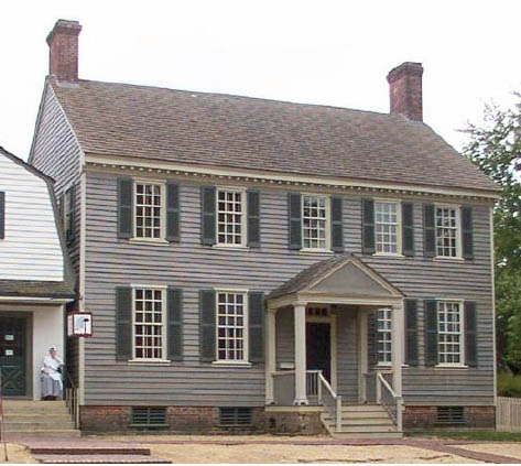 Charlton House, Block 9, Building 30, Lot 22
| Structure: | Charlton House, Block 9, Building 30, Lot 22 |
|---|---|
| Requested by: | Edward A. Chappell, Roberts Director of Architectural and Archaeological Research, Colonial Williamsburg Foundation |
| Analyst: | Kirsten Travers, Graduate Fellow, Winterthur/University of Delaware Program in Art Conservation |
| Supervised by: | Susan L. Buck, Ph.D., Conservator and Paint Analyst, Williamsburg, VA |
| Date Submitted: | December 15, 2010 |
Purpose:
The goal of this project is to use cross-section microscopy, fluorochrome staining and polarized light microscopy techniques to identify and characterize any surviving early paints on the Charlton House exterior, particularly those that may date to the late-eighteenth century. The results will be used to design a new, more historically accurate color scheme for the building.
Historical Background:
There is some debate concerning the construction and subsequent alterations of the Charlton House (dendrochronological dating has not been carried out). As recently as December 2010, stylistic analysis by Edward Chappell (Director of Architectural and Archaeological Research), suggests that the cornice could date to the post-Revolutionary period. He explains that the combination of delicate dentils and modillions on the cornice of a house of this scale became more common after the 1780s. However, he also asserts that other parts of the house are convincingly pre-Revolutionary, in particular the door leaves and doweled flooring. Furthermore, it appears that the 1929-30 restoration adjusted the early 19th-century state of the house, because early wood framing appeared to have been reused in the restored roof (which contained cut nails (post-1800), as well as wrought nails). In other words, Chappell suggests that in the restoration period, the handsome exterior cornice and beaded siding that was retained was early, but may not be first-period.
Previous research based on documentary evidence suggests that Edward Charlton, a wigmaker and barber, purchased lot #22 as early as 1772, and may have occupied a house on the property by 1774 (Stephenson 1957, 8). Preparing to leave the colonies for England, Edward Charlton and his wife Jane, a milliner, advertised their house for sale one three separate occasions: in 1774, 1775, and 1778, but it is not known if this is the house currently situated in lot 22.
4Their latest advertisement, dated May 22, 1778, reads:
To be rented or sold, the HOUSE and LOT…belonging to the subscriber, situate on the most public part of the main street…well calculated for any business, and in good repair (Stephenson 1957, 10).
The Charltons were unable to sell their house, and a deed dated 1784 transferred the building to the estate of his deceased brother, Richard Charlton, in that year. Richard Charlton's widow and children lived in the house until 1795, when it was conveyed to William Russell, a Williamsburg clerk. The next year Russell insured the property, which he described as:
My wooden buildings on the main street at Williamsburg…a dwelling house of wood— two stories 44' by 29', office of wood— one story 30' by 26' and a kitchen of wood— one story 22' by 18'…on the lot (Stephenson 1957, 12).
Russell lived in the house on lot 22 until his death, and in 1816 it was conveyed to his daughter, Catherine. From 1819 to 1880 the house was owned by the Henley family, and during this time it became known as the "Old Henley Place." In the late 19th century, the house was purchased and occupied by the Servant family. In 1928, Mary Lucy Servant [Servient] conveyed the property to W.A.R. Goodwin (Stephenson 1957, 20).
Colonial Williamsburg's reconstruction of Charlton House (then referred to as the Servient House), began in October 1929 and was completed six months later (Kocher and Dearstyne 1950). During that time it was discovered that the house has been altered significantly from its original appearance. The Charlton House Architectural Report states that:
The house depth had been reduced to about ½ of its original dimension; the house had at some time literally been cut in two along its east-west axis for the full length of the building causing demolition of all original chimneys. This was evident upon the discovery and study of the old foundation walls in the basement, which gave to the restorers the exact dimensions of the original building and also the location of the chimneys (Kocher and Dearstyne 1950, 21).
During the restoration, all 19th-century additions and alterations were removed, and "old" work (denoting anything that cannot be defined with certainty as being original but which is old enough to justify its retention in a restored building), was retained and repaired. The architectural report lists the "old" exterior elements as:
…the front cornice, the weatherboarding on the north side and some on the west, the corner boards on the northeast and northwest corners, exterior trim and sills of second floor windows of the north front, frames and sills of the end basement windows of the north front…(Kocher and Dearstyne 1950, 32).5
Exterior color: Black and white photographs of the house before restoration show that c. 1929, the north cornice was painted a much darker color than the weatherboards (Kocher and Dearstyne 1950). According to CWF paint shop records, the house has been painted nine times since 1930. The same colors have been repeatedly applied since the restoration (see Table 1):
| Element | Color | Notes |
|---|---|---|
| Weatherboards | Peyton-Randolph House gray | CW commercial color #1086 |
| Exterior Trim (including cornice) | Light gray | CW custom color #733 |
| Shutters, window trim | Dark green | CW custom color #1 |
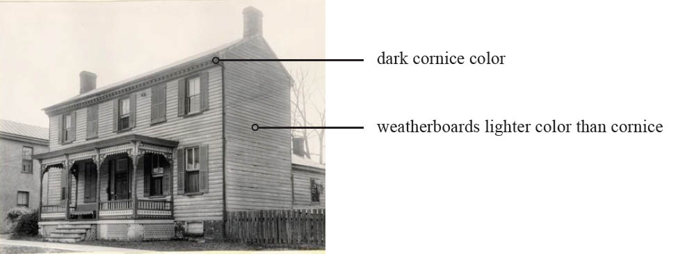 Charlton House, northwest view before restoration, c.1929 (CWF archives)
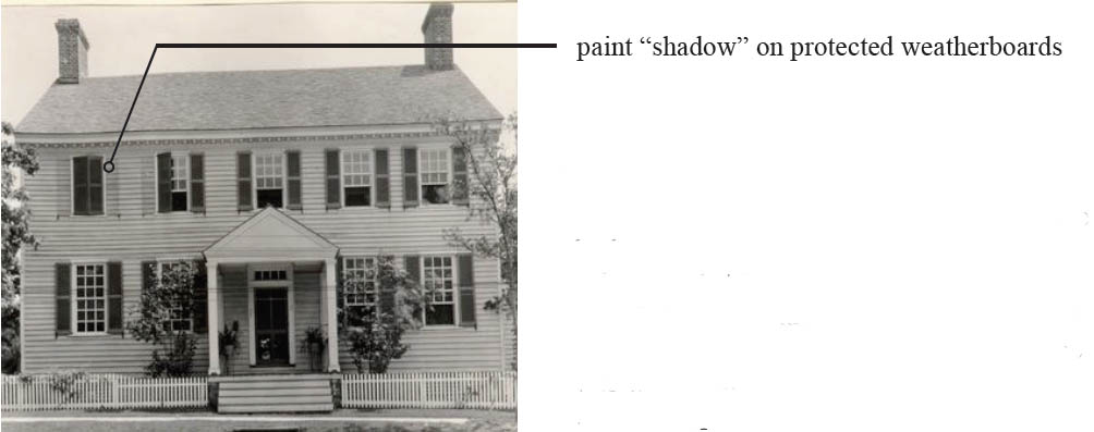 Charlton House, northwest view after restoration, precise date unknown (CWF archives)
The photograph of the house after restoration was included in the 1950 Kocher and Dearstyne Charlton House Architectural Report. Susan Buck noted that the shadows on the weatherboards behind the shutters where the paint was protected from light exposure suggests that this photo was taken some time after the restoration (Buck 2010, pers. comm.).
Previous Finishes Research:
In 2002, Catherine Matsen used cross-section microscopy techniques to determine the paint history of a weatherboard fragment from the Charlton House in the architectural fragments collection (Matsen 2002). She identified six (6) finish generations, starting with a first generation white paint disrupted by dirt and mold. The second paint generation is white, while the third is a cream color that appears to have a varnish component, possibly added increase durability and gloss to the final finish. Generation four is a gray color, generation five a white color, and generation six is a gray color — the current presentation surface. Binding media analysis was not carried out as part of her research.
Procedures:
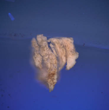
Sampling: In 1996, Tom Taylor (former CWF Director of Architectural Conservation), removed four samples of wood and paint from the Charlton House exterior north cornice. On average, the wood samples measured ¾" long and ¼" wide and retained significant finish evidence. Each sample location was noted (written documentation only), and the samples were stored in labeled plastic baggies that were incorporated into the architectural fragments collection as individual objects (CHH-[004-007]-08).
On July 20, 2010, Natasha Loeblich (CWF paint analyst), removed four smaller paint samples from Taylor's 1996 cornice fragments, and also collected three samples from the original Charlton House weatherboard in the collection (CHH-002-08). Where applicable, the sample sites were recorded with written documentation.
On July 21, 2010, fourteen samples were collected from original exterior elements on the Charlton house exterior by Natasha Loeblich and Edward Chappell (CWF Director of Architectural and Archaeological Research). The sample locations were recorded with written and photographic documentation, and the samples were stored in individual labeled plastic bags and transported back to the Susan Buck's laboratory for analysis.
Laboratory: The paint samples were cast and polished by Natasha Loeblich in the summer of 2010. The microscopy and fluorochrome staining was carried out by Kirsten Travers (CWF graduate intern) in the fall of 2010.
The uncast samples were examined with a stereomicroscope under low power magnification (5x to 50x), to identify those that contained the most paint evidence and would therefore be the best candidates for cross-section microscopy. Uncast sample portions were retained for future examination and analysis. The best candidates were cast in resin cubes and sanded and polished to expose the cross-section surfaces for microscopic examination. Please see Appendix A for sample preparation details.
All of the cross-section samples were examined and digitally photographed in reflected visible and ultraviolet light conditions at 20x to100x magnifications. Important visual characteristics 7 were recorded at this stage, including layer color and thickness, the level of surface deterioration, the presence of dirt layers, and autofluorescence in reflected ultraviolet light.
Results:
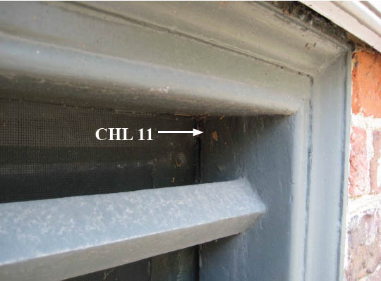
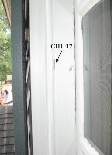
In all of the samples, the earliest paint layers are extremely disrupted due to age, weathering, and biological growth. However, some samples from the cornice and weatherboards retain adequate paint evidence to interpret the first generation color scheme for these elements. The samples removed from the cornerboards do not appear to contain historic paints, and are discussed briefly. The samples from the second-floor window frame and basement window grill contain modern paints only and were not interpreted for this report.
The most relevant results are presented in the following section. For clarity, the data has been organized by element, ie: cornice, weatherboards, and cornerboards. Each section summarizes the results for that element, illustrated by the most informative photomicrographs with corresponding annotations and comments from the author. The results are interpreted in the conclusion. All raw photomicrographs can be found at the end of this report.
Cornice:
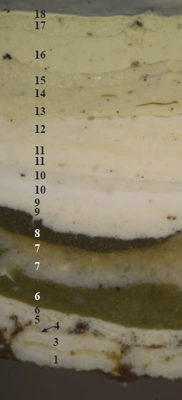
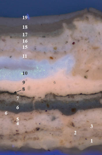
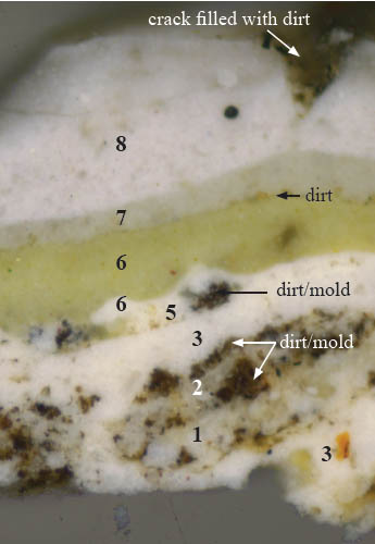
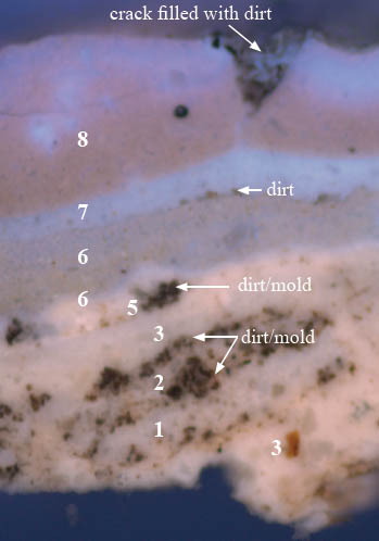
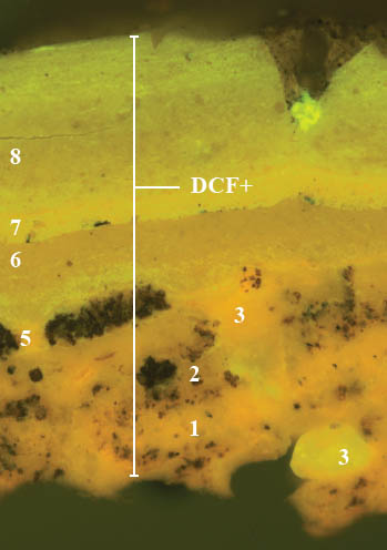
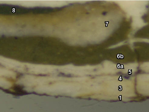
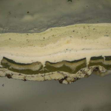
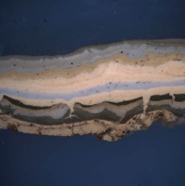
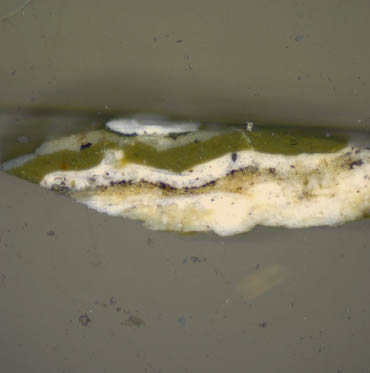
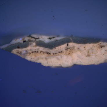
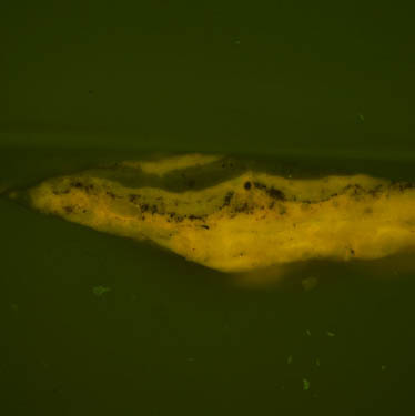
Cornice samples CHL 3, CHL 9, and CHL 10 retained the most intact paint evidence from which the decorative history of the cornice could be studied. Please note that primers and finish coats are noted as the same generations in the following annotated photomicrographs.
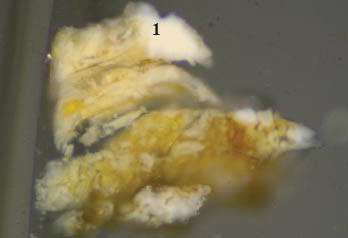
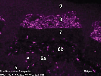
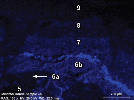
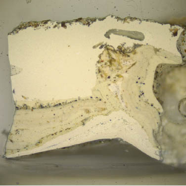
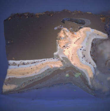
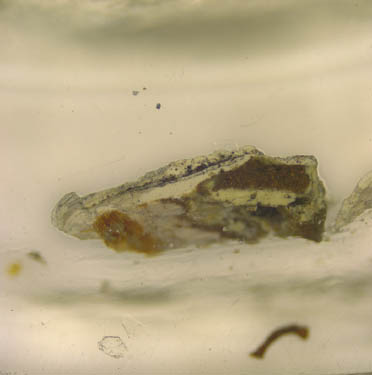
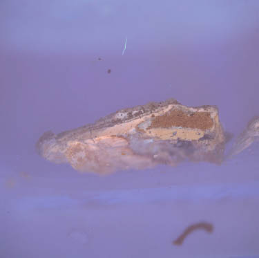
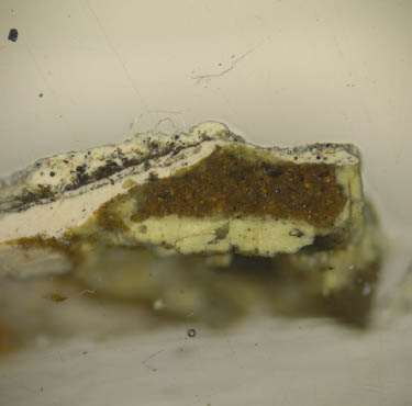
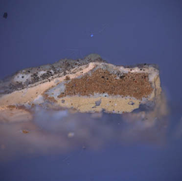
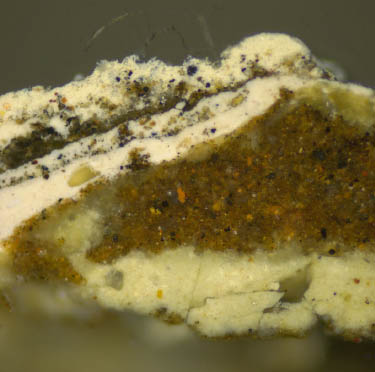
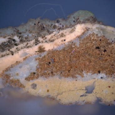
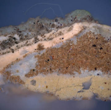
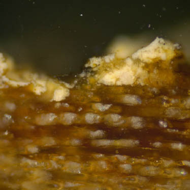
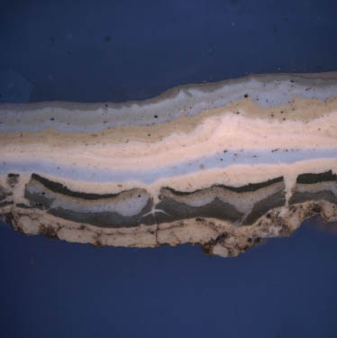
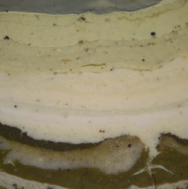
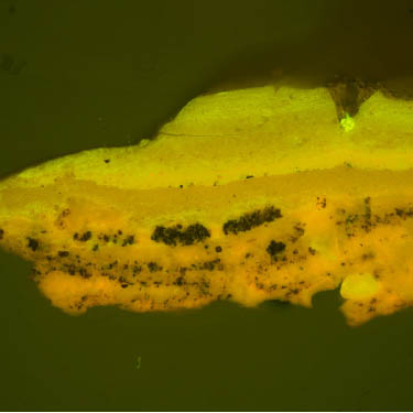
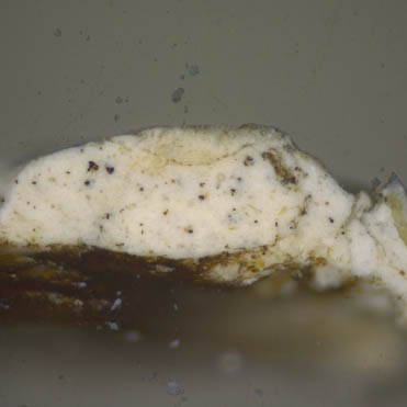
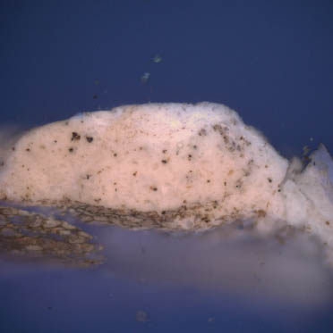
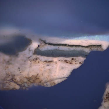
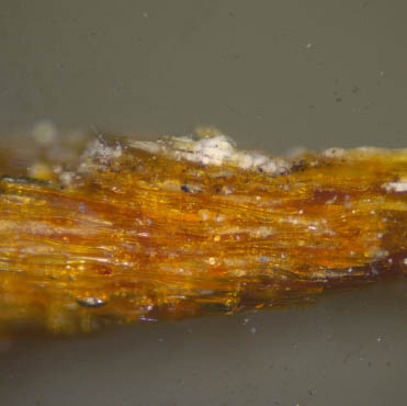
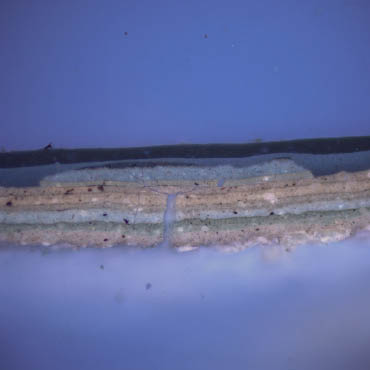
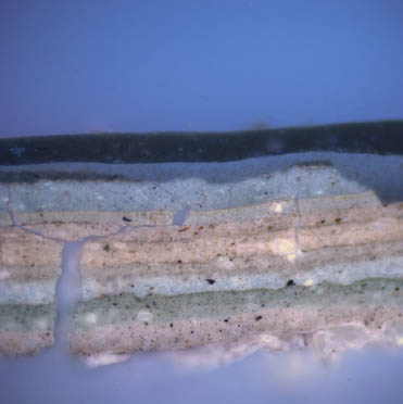
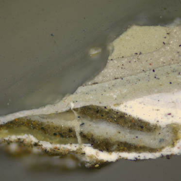
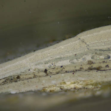
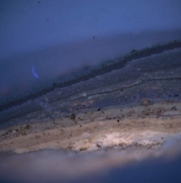
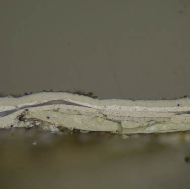
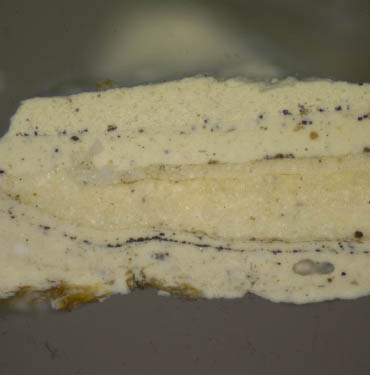
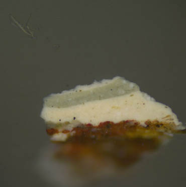
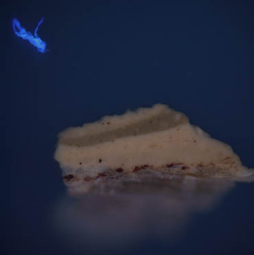
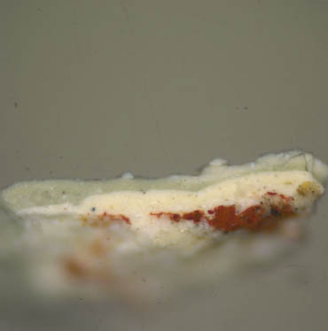
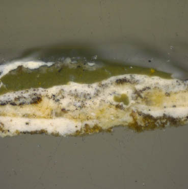
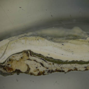
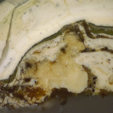
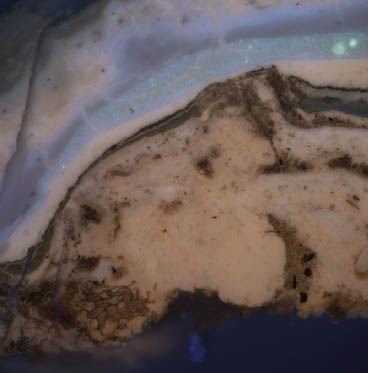
Sample CHL 3b: Cornice fragment CHH-006-08, removed from bed molding around the modillion blocks just before the house was repainted in 1996.
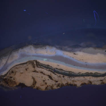
 CHL 3b, visible light, 100x
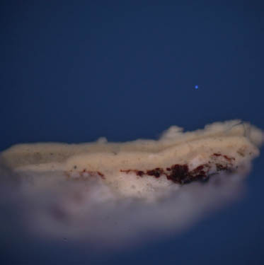
 CHL 3b, UV light, 100x
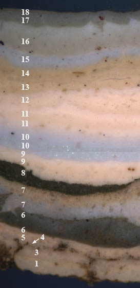
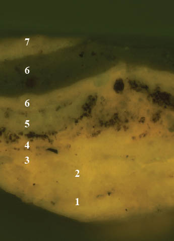
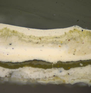
Sample CHL3b is missing generation 2, but otherwise retains the most complete stratigraphy in the sample set. The first five finish generations are white paints that display a dim, pinkish autofluorescence suggestive of white lead in a drying oil (although this would need to be confirmed with an instrumental technique such as scanning electron microscopy-energy dispersive spectroscopy (SEM-EDS), x-ray diffraction (XRD), or Raman spectroscopy). Generation 1 is a cream-colored paint with large, coarsely ground white particles. The distinct boundary between generations 1 and 3 indicates that generation 1 had dried completely before the next was applied, suggesting that it was indeed a presentation surface.
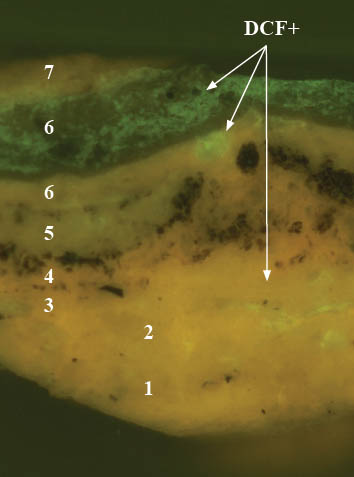
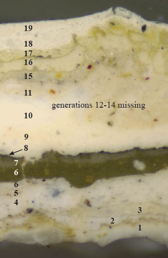
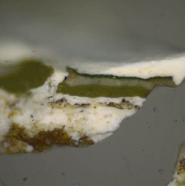
Generation 4 is a highly degraded white paint layer which is heavily soiled and appears to have been attacked by mold, suggesting an extended period of exposure and neglect. The dirt and mold appear to have disrupted the paints and extend down to the substrate during this period, after which it was painted with generation 5, another white paint. The sixth paint generation consists of two layers, a white base coat and olive-green top coat. This is followed by generation 7, a gray paint with a dim autofluorescence at its surface, suggesting exposure and wear. Generation 8 is a dark green paint that exhibits a cracked and deteriorated surface, seen also in cornice samples CHL 9 and CHL 10. This green could be the darker color on the cornice in the c.1929 photograph of the house prior to restoration.
All generations applied after this dark green paint (generations 9 through 18), are the primers and off-white finish coats applied by CW since 1930. The particles in generations 10 and 15 display the "twinkling" bright blue autofluorescence that is characteristic for zinc white pigment (introduced c.1840), although this would need to be confirmed with instrumental analysis (see above).
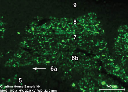
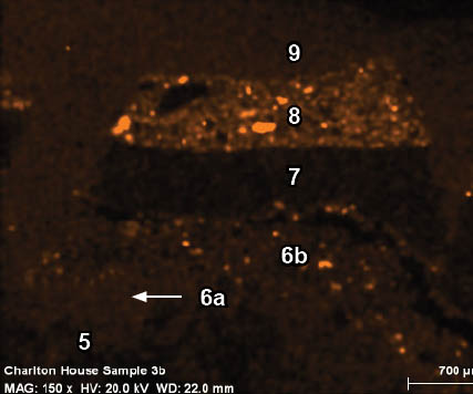
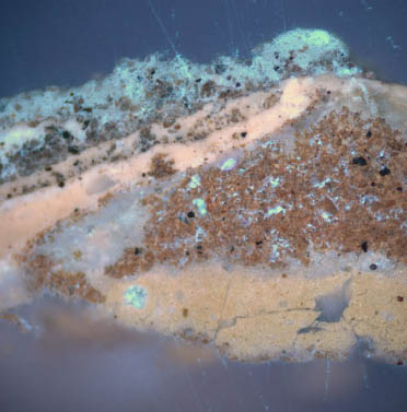
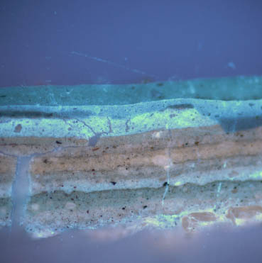
Fluorochrome staining results:
 CH 3b, UV, 100x. Before TSQ.
 CH 3b, UV, 100x. TSQ reaction.
The sample was stained with TSQ to detect the presence of Zn+2, possibly present in zinc white, a pigment available only after c.1840. Reactions were observed in generations 6, 7, 10, and 15-18. No reactions were observed in the earliest paint generations.
10 CHL 3a, visible light, 200x
Wood substrate and early paint.
 CHL 3a, UV light, 200x
Wood substrate and early paint.
Two early paint layers are attached to the cornice wood substrate. Generation 1 appears to be the cream-colored paint observed in other cornice samples CHL 3b and CHL 10. Generation 2 is the white paint that was missing from sample CHL 3b. This layer is less yellow and has a slightly brighter autofluorescence in comparison to generation 1. However, this evidence is only very fragmentary.
Fluorochrome staining results:
 CHL 3a, UV light, 200x, before TSQ.
 CHL 3a, UV light, 200x. TSQ reaction.
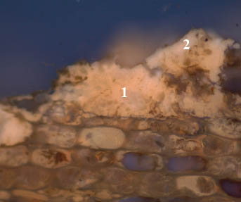
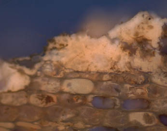
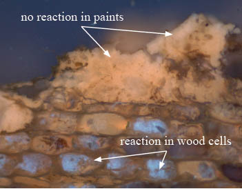
Fluorochrome staining with TSQ of sample CHL 3a determined that Zn+2 was not present in the early paints. However, the characteristic reaction for the free zinc ion- a bright blue autofluorescence, was observed in the wood cells. It is possible that this is a false positive, or represents a later zinc-containing paint that was applied when the surface had weathered to bare wood, and flowed into the wood substrate.
11 CHL 3a, B2A filter, 400x. Before DCF.
(TSQ reaction still visible in wood cells).
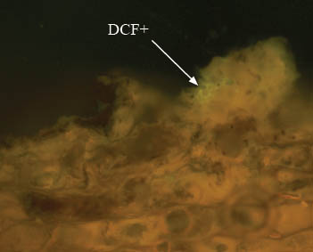 CHL 3a, B-2A filter, 400x. DCF reaction.
(TSQ reaction still visible in wood cells).
Sample CHL 3a was stained with DCF to tag for the presence of oils. There was a minor reaction (a bright yellow-green fluorescence), in the earliest paint layers, suggesting that lipids, ie: oils, are present. The weakness of this reaction could result from the age and deterioration of the oil binding media.
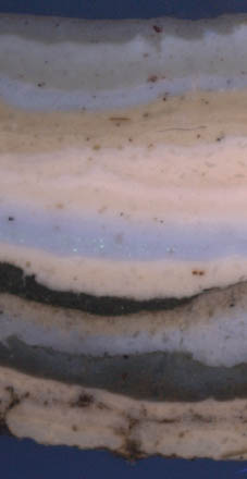
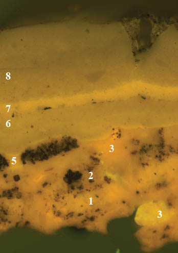
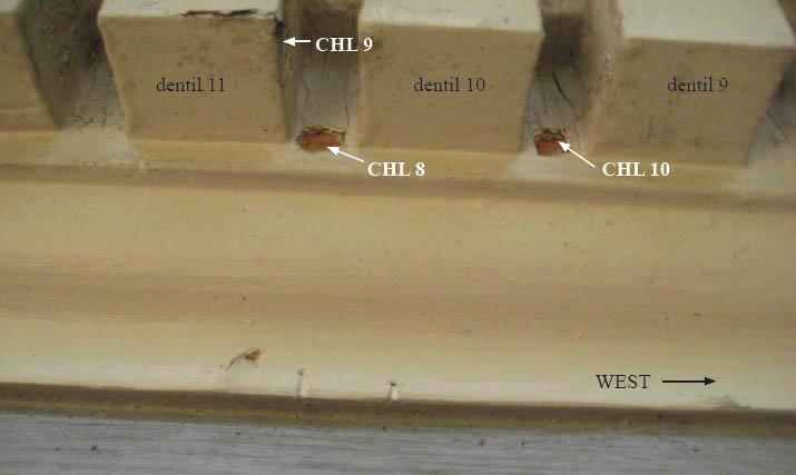
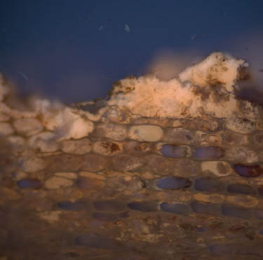
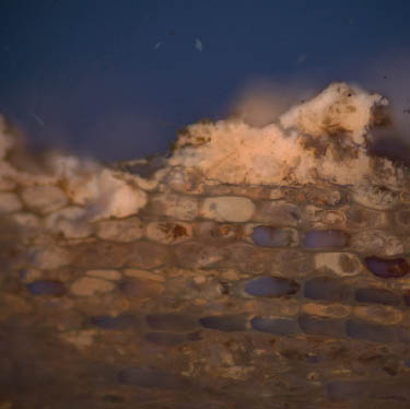
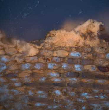
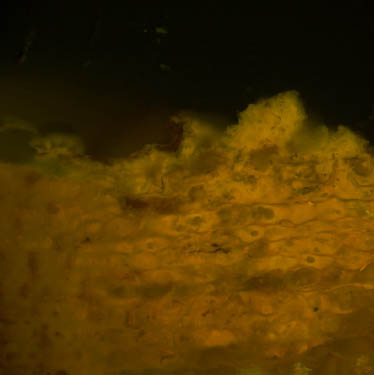
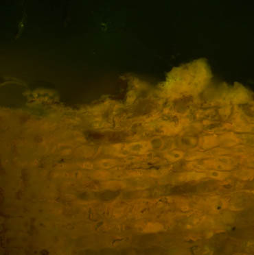
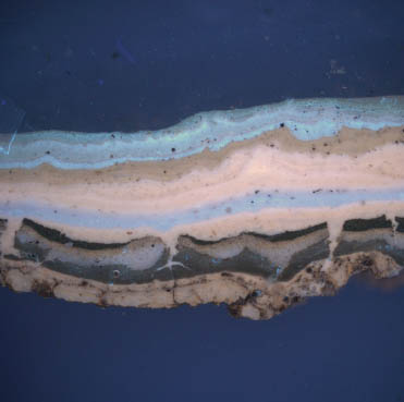
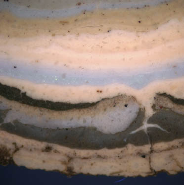
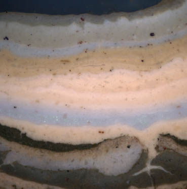
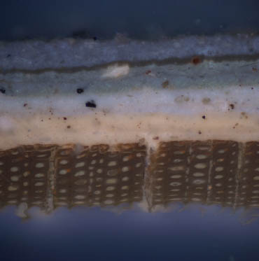
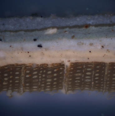
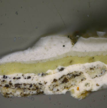
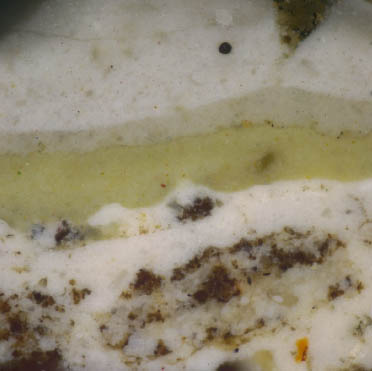
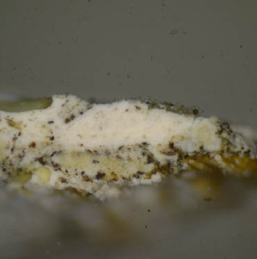
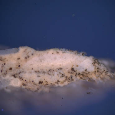
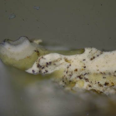
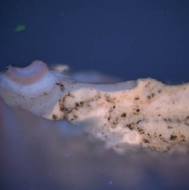
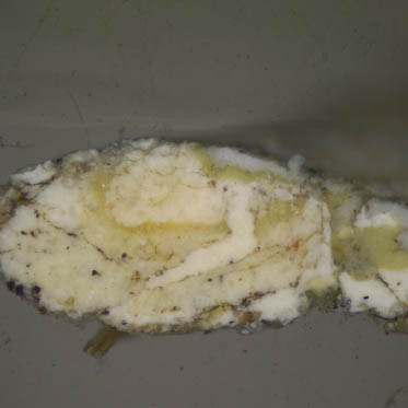
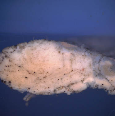
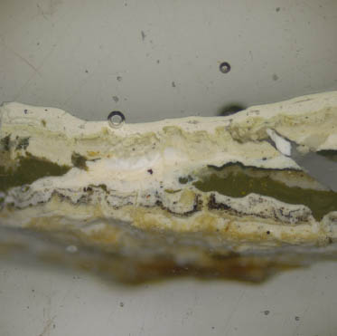
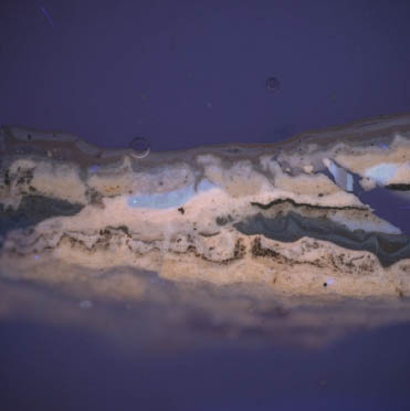
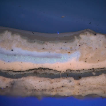
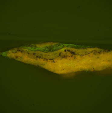
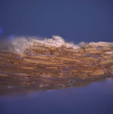
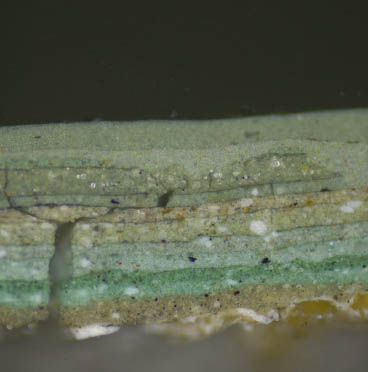
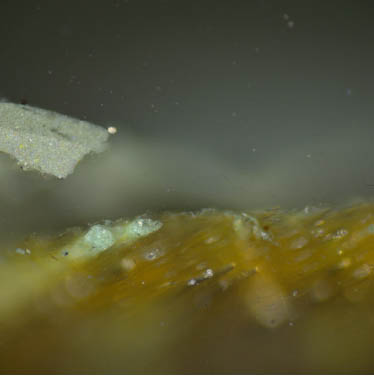
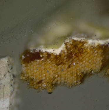
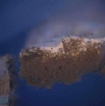
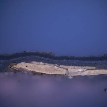
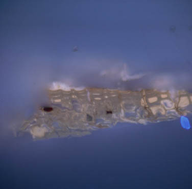
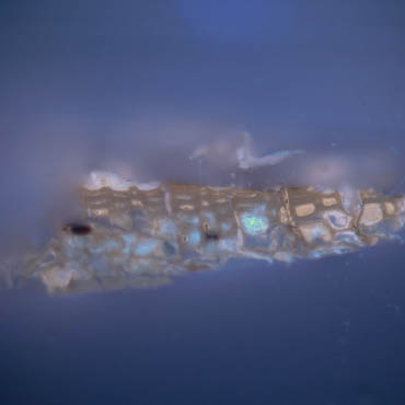
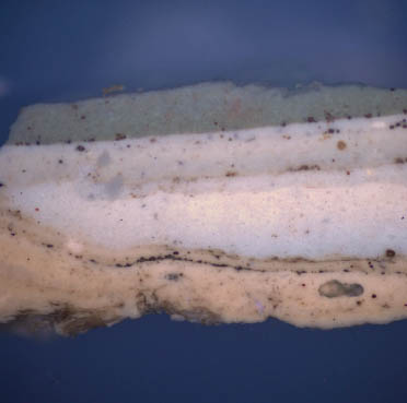
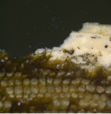
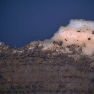
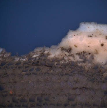
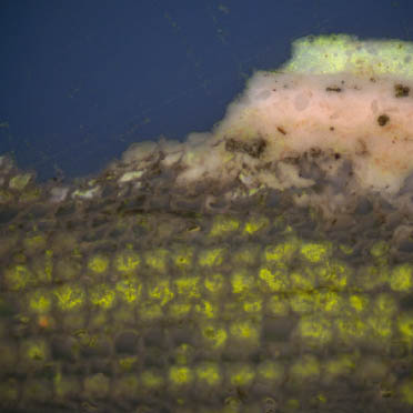
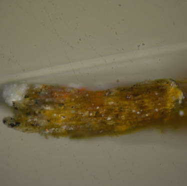
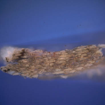
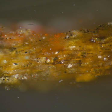
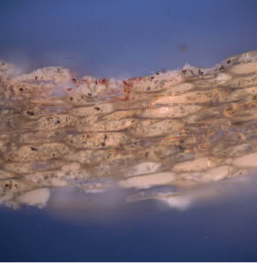
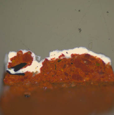
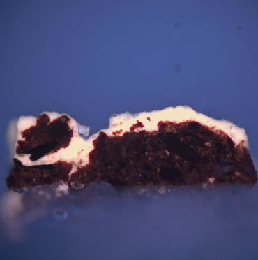
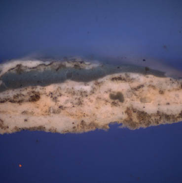
Sample CHL 10: North elevation, cornice fascia between dentils 10 and 11 from the west end.
 CHL 10, visible light, 100x (enlarged)
 CHL 10, UV light, 100x (enlarged)
 CHL 10, visible light, 100x (wood substrate and early paint)
 CHL 10, UV light, 100x (wood substrate and early paint)
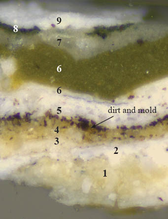
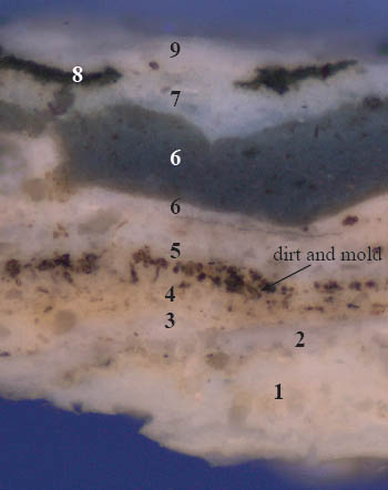
This sample contains the most intact early stratigraphy. Generation 1 exhibits large, coarsely ground white particles in a yellowish binding medium- possibly an oxidized drying oil. A thin film of dirt is visible on the surface of this paint, suggesting that it was a presentation surface. Generations 2-5 are various shades of white and off-white, distinguished by their autofluorescence colors and the layers of dirt, mold, and grime which separate them.
13The large amount of mold on the surface of generation 4 suggests this surface was exposed for an extended period of time.
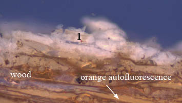
The earliest paint generation attached to the wood substrate associated with sample CHL 10 is a degraded white paint. The orange-colored autofluorescence in the wood fibers suggests that shellac was used to seal the surface before painting.
Fluorochrome staining results:
 CHL 10, B2A filter, 100x (enlarged). Before DCF.
 CHL 10, B2A filter, 100x (enlarged). DCF reaction.
Sample CHL 3a was stained with DCF to tag for the presence of lipids (oils). Weak and 'blotchy' positive reactions (a bright yellow-green fluorescence), were observed in generations 1 and 2, suggesting they are oil-bound. The weakness of these reactions could be the result of the extensive deterioration of the binding media in these early paints. Stronger reactions were observed in generations 5-7.
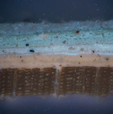
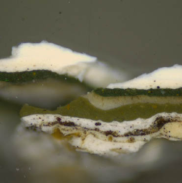
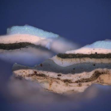
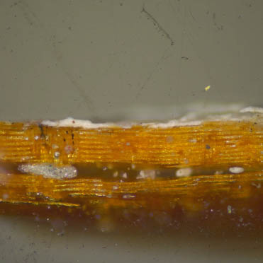
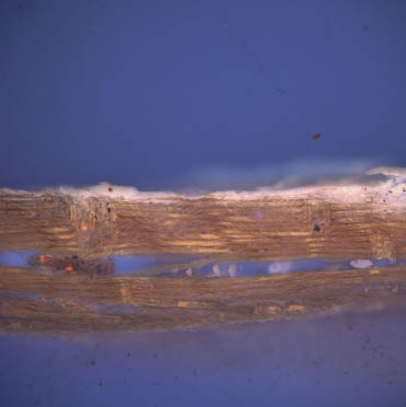
Sample CHL 9: North elevation, cornice, west side of 11th dentil from west end
 CHL 9, visible light, 100x
 CHL 9, UV light, 100x
 CHL 9, visible light, 100x
Wood substrate and trapped early paints
 CHL 9, UV light, 100x
Wood substrate and trapped early paints
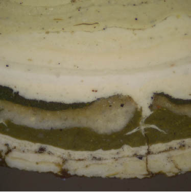
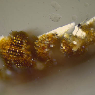
Sample CHL 9 contains a similar stratigraphy to the previous cornice samples. The first paint generation is a cream-colored paint with a pinkish autofluorescence. The thin layer of dirt on the top of this layer suggests that it was a presentation surface. Generations 2-5 are white and off-white paints separated by layers of dirt and grime. Generation 6 is comprised of a white primer with an olive green-colored finish coat. Generation 7 is a gray paint. This is followed by generation 8, one layer of dark green paint. This generation is much thicker in sample CHL3b (page 8). The cracks in the dark green paint suggest it was exposed for an extended period of time.
All layers applied after this dark green paint (generations 9 to 19), may be the primers and off-white finish coats applied by CW since 1930. Generations 12-14 are missing from this sample, probably lost during routine sanding and/or scraping prior to re-painting. The particles in generations 10 and 15 display the "twinkling" bright blue autofluorescence that is suggestive of zinc-based pigments, the earliest of which is zinc oxide(ZnO, introduced c.1840), but this would need to be confirmed with instrumental analyses.
The earliest paint layer attached to the wood substrate associated with sample CHL 9 is a white paint. The orange-colored autofluorescence in the wood fibers suggests that shellac was used to seal the surface before painting. However, the presence of shellac would have to be confirmed with other analytical methods, such as FTIR (Fourier transform infrared spectroscopy), or GCMS (gas chromatography-mass spectrometry).
Cornice, Polarized Light Microscopy results:
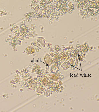
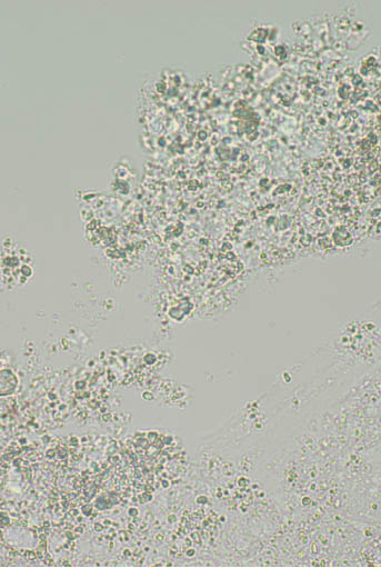
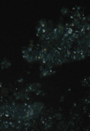
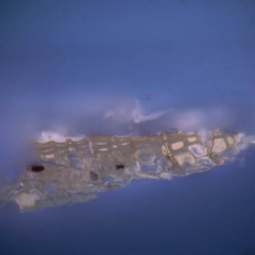
Fragments of paint layer 1 were found deep in the fibers of the uncast wood substrate from the cornice fragment. Scrapings from these remnants were analyzed with polarized light microscopy, and found to contain lead white (2PbCO3 ∙ Pb(OH)2), and large particles of chalk (Ca CO3), which has little hiding power in oil, but is commonly added as an extender in house paints (Gettens and Stout 1942, 104).
 Scraping from cornice fragment, generation 1 paint.
Transmitted plane polarized light, 1000x
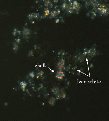 Scraping from cornice fragment, generation 1 paint.
Transmitted cross polarized light, 1000x
Weatherboard:
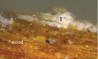
None of the weatherboard samples contain a complete paint history. However, sample CHL 5a retains the most intact early paints, and is discussed in the following section. The remaining samples from the weatherboard fragment (CHL 6, 7) retain early paints that were deteriorated to such an extent that interpretation was not possible. The samples removed in-situ (CHL 12, 13, 19) retain only modern paint layers and are not discussed in this report.
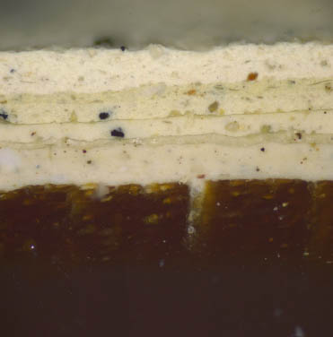
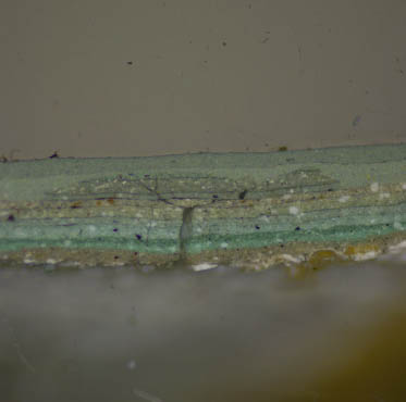
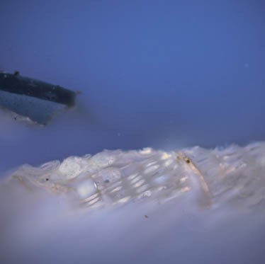
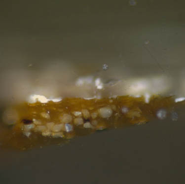
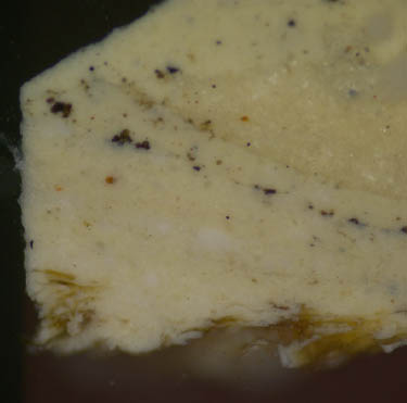
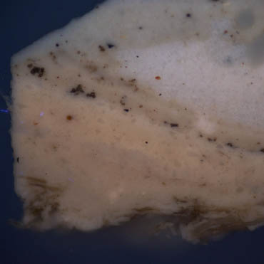
Sample CHL 5a: Weatherboard fragment CHH-002-08
 CHL 5a, visible light, 100x
 CHL 5a, UV light, 100x
The early paint layers in this sample are very degraded and compromised by dirt and mold. However, adequate evidence remains to make relevant comparisons. Generation 1 is a coarsely ground paint made with white particles in a yellowish-matrix. This layer has a pinkish-autofluorescence in reflected ultraviolet light. These characteristics are similar to the first white paint layer on the cornice (see sample CHL 3), suggesting that these layers are contemporary, and that in the first finish generation, the cornice and weatherboards were painted white.
Generations 1 and 2 appear to have deteriorated to such an extent that they detached from the wood substrate, allowing generation 3 to flow around and encapsulate them during application.
18Generation 4, the severely dirty and deteriorated white paint generation seen in the cornice samples, is absent here. Generation 5 is comprised of a white primer with a dull yellow-green finish color that has a thin layer of grime on its surface. In the next finish generation (7), the weatherboards were painted gray, followed by generation 8, a light gray finish coat. This light gray layer exhibits large cracks in its surface that have begun to fill up with dirt, suggesting a long period of exposure.
Fluorochrome staining results
 CHL 5a, B2A filter, 100x. Before DCF.
 CHL 5a, B2A filter, 100x. DCF reaction.
Sample CHL 5a with was stained with DCF to tag for the presence of lipids (oils). Reactions were observed in all layers. Strong reactions (a yellow-green fluorescence), were observed in generations 3 and 5-8. Weak, 'blotchy' reactions were observed in generations 1 and 2. This could result from the age and deterioration of these layers, resulting in leaching of the binding media.
Weatherboard, Polarized Light Microscopy Results:
Fragments of paint generation 1 were found deep in the fibers of the weatherboard fragment. Scrapings from these remnants were examined in transmitted plane and cross polarized light, and found to contain lead white (2PbCO3∙Pb(OH)2), and large particles of chalk (Ca CO3). Chalk has little hiding power in oil, but is commonly added as an extender in house paints (Gettens and Stout 1942, 104). These pigments are the same as those identified on the cornice.
 Scraping from weatherboard fragment.
Transmitted plane polarized light, 1000x.
 Scraping from weatherboard fragment.
Transmitted cross polarized light, 1000x.
Cornerboard:
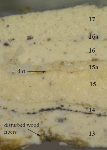
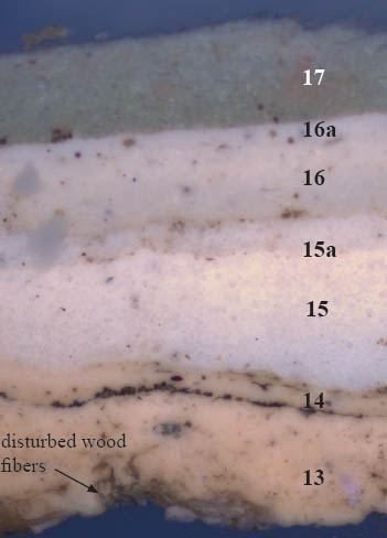
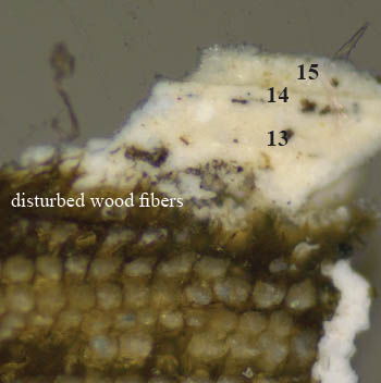
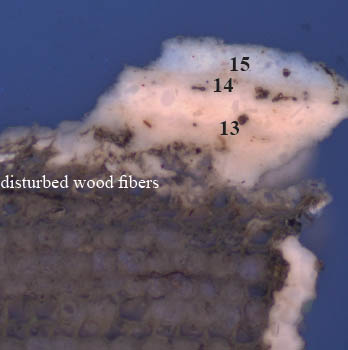
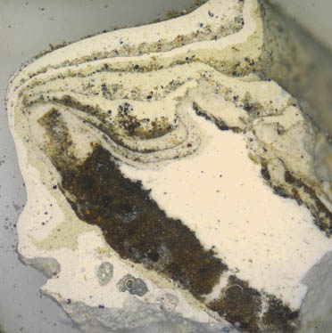
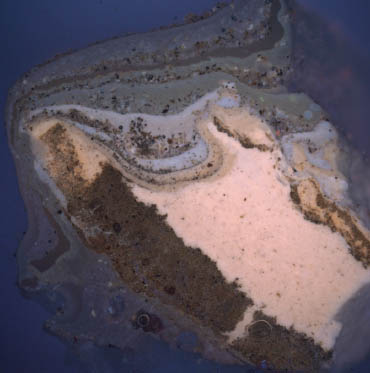
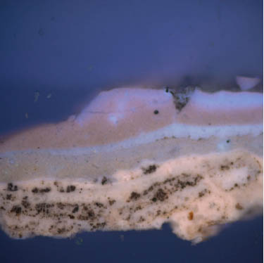
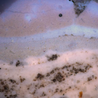
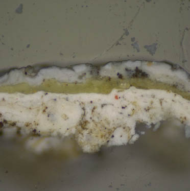
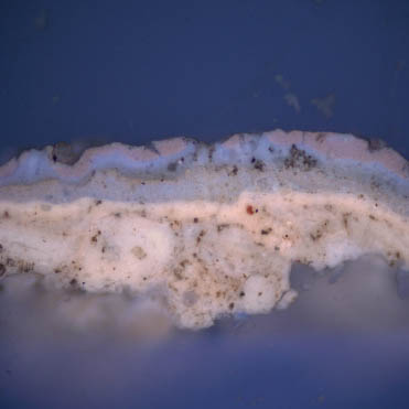
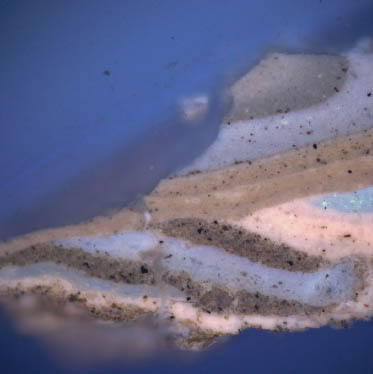
The cornerboard samples do not appear to contain any of the earliest paints. In sample CHL 14, only seven generations are extant, most of which can be lined up with the later 20th-century paints on the cornice (see sample CHL 3b, page 8). Generations 13 and 14 display a pinkish-autofluorescence characteristic for white lead in oil, but the finely ground particles suggest an industrially prepared paint. In addition, the substrate fibers trapped in the paint at the bottom of the sample suggests that the surface was sanded or scraped at some point in its history, removing the previous paint evidence.
Generations 15 and 16 are the same as those seen in other exterior samples, however, these paints appear to have been re-applied after a certain period of exposure (note the thin dirt layer separating layer 15 from layer 15a). This pattern was not observed in all samples, and would suggest that the cornerboards were "touched up" where necessary by the CW paint crew.
 CHL 14, visible light, 100x
 CHL 14, UV light, 100x
 CHL 14, visible light, 200x.
Wood substrate and paint layers.
 CHL 14, UV light, 200x.
Wood substrate and paint layers.
Color Measurement Results
Color measurements were attempted on the first generation white paint trapped in the wood fibers of the weatherboard and cornice fragments. However, after several attempts it was clear that due to of the small amount of paint remaining, its extreme deterioration, uneven yellowing, and embedded mold and soiling materials, it would be impossible to obtain an accurate color reading. Since the analysis determined that the first generation paint was made with lead white and chalk in linseed oil, the first generation paint remnants were compared by eye (under 50x magnification with a color corrected light source), to an actual "mock-up" sample of white paint made with lead white in linseed oil in the paint analysis laboratory (this paint sample was hand-made by Susan Buck, Ph.D., in 2004 and painted out on a shellacked pine board). The comparison found that the color and texture of the 2004 lead white paint sample matched very well with the remnants of first generation Charlton House paint. Therefore, the 2004 lead white painted board was measured with the Minolta Chroma Meter to provide substitute color values for the Charlton house first generation white paint (for additional information on color measurement procedures, see Appendix B). Three measurements in CIE L*a*b* colorspace values were taken from the 2004 white paint sample, and the average was calculated. The CIE L*a*b* values can be found in the table below:
| Color Values | L* | a* | b* |
|---|---|---|---|
| Measurement 1 | 98.59 | -0.23 | +1.84 |
| Measurement 2 | 99.08 | -0.05 | +0.80 |
| Measurement 3 | 98.71 | -0.03 | +1.18 |
| Average | 98.79 | -0.10 | +1.27 |
Using a stereomicroscope at 30x magnification with a color corrected light source, the Charlton
House paint remnants and the 2004 lead white paint mock-up were compared by eye to various
commercial color swatches to provide a match for the first generation paint. The closest swatch is
attached below:
 Benjamin Moore #OC-33 "Opaline"
| Color value | L* | a* | b* |
|---|---|---|---|
| Measurement 1 | 98.59 | -0.23 | +1.84 |
| Measurement 2 | 98.76 | -0.28 | +1.77 |
| Measurement 3 | 98.88 | -0.26 | +1.75 |
| Average | 98.74 | -0.26 | +1.79 |
Using the equation found on page 31, the color difference (ΔE), between the white lead paint sample and the commercial swatch above was determined to be 0.5. Since the human eye cannot detect color differences with a ΔE value of 3 or below, Benjamin Moore color OC-33 is therefore an excellent color match for the lead white paint mock-up.
Conclusions:
Of the 21 samples taken from Charlton House, few retain the earliest paints, but cornice samples CHL 3a, CHL 3b, CHL 9, and CHL 10, and weatherboard sample CHL 5a did contain enough evidence to reconstruct the first generation paint scheme that had been applied to those elements. In all of the samples, the earliest layers were soiled, weathered, and fragmented, which is typical for paints exposed to outdoor environments for an extended period of time. In addition, many of the CW house painting records relate that the Charlton house has been sanded, washed, and scraped in the past, which would have further contributed to the loss of early paint histories. This study found that samples from the cornerboards, window frames and basement window grilles did not retain any historic paint evidence, so their earliest appearance could not be determined. However, analysis of the surviving paints suggest that the early Charlton House color scheme was very different from how it appears today.
The results suggest that the wood surfaces of Charlton House were first sealed with a resinous sealant, possibly shellac, prior to painting. However, the presence of shellac must be confirmed with further analysis using an instrumental technique, such as FTIR (Fourier-transform infrared spectroscopy), or GC-MS (gas chromatography-mass spectrometry). On the cornice and weatherboards, the first finish generation is a white oil-bound paint made with chalk (CaCO3), and lead white (2PbCO3 ∙ Pb(OH)2) pigments. In the uncast sample, the remnants of this white paint appear light yellowish-gray with black pigments embedded in it. Since no colored pigments were visible in the numerous cross-sections or in the dispersed pigment samples taken from this layer, it seems likely that these particles are not pigments, but soils that became embedded in the paint over time as the layer deteriorated. The yellowish cast most likely results from the oxidation of the drying oil medium. Several attempts were made to obtain colorimetry measurements for the first generation white paint, but its deterioration made it impossible to obtain an accurate color reading. Instead, an approximate color match was based on a mock-up sample white paint made with lead white pigment in linseed oil. To recreate the look of this traditional paint on the Charlton House exterior, a commercial color match is provided on page 23.
The identification of a first generation white paint made with lead white and chalk is consistent with an 18th-century description of Williamsburg, written in 1783 by Johan Schoepf, a surgeon to the British Army: "the houses stand at convenient distances apart, have a good exterior, and on account of the white paint, a neat look" (Rowe 1983, 14).
The composition of this first white paint as containing a mixture of white lead and chalk is also consistent with John Lord Sheffield's 1784 account in Observations on the Commerce of the American States: "chalk and white lead form at least three-fourths of all paint" (Rowe 1983, 15).
Following the initial white paint scheme, the exterior appears to have been painted white repeatedly for up to four generations. The samples from the weatherboards and cornice can be aligned to suggest that in the 6th generation, the weatherboards were painted a dull yellowish-green while the cornice was painted an olive green. The physical characteristics of these paints suggest that this was a late 19th, or early 20th-century paint scheme.
25In the next paint generation, both the weatherboards and cornice were painted gray. Following this, in generation 8, the weatherboards were painted light gray while the cornice was painted a dark green color. The top surface of generation 8 is cracked and soiled, suggesting that it was exposed for a very long period of time. This is believed to be the immediate pre-restoration appearance of the house, which black and white archival photographs show as having dark trim with lighter-colored weatherboards.
Since the 1930 restoration, Charlton house has been repainted approximately once a decade. The color scheme consists of gray weatherboards, light gray trim, and dark green shutters. These gray and light gray paints and their associated primers are visible as generations 8-18.
References
- Gettens, R., and G. Stout. 1942. Painting materials: a short encyclopedia. New York, Dover Publications, Inc.
- Eastaugh, N., et. al. 2008. Pigment Compendium: a dictionary and optical microscopy of historical pigments. Oxford, Butterworth-Heinemann.
- Matsen, C. 2002. Cross-section microscopy report: architectural fragments in the Colonial Williamsburg collection. Unpublished report for the Architectural Research Department, Colonial Williamsburg Foundation,Virginia.
- Kocher, A. and H. Dearstyne, 1950. Charlton House Architectural Report, Block 9, Building 30, Lot 22. Unpublished report for the Architectural Research Department, Colonial Williamsburg Foundation, Virginia.
- Rowe, L. 1983. Exterior Painting. Unpublished report for the Colonial Williamsburg Foundation, Virginia.
- Stephenson, M. 1957. Charlton House Historical Report, Block 9, Building 30, Lot 22. Unpublished report for the Historical Research Department, Colonial Williamsburg Foundation, Virginia.
- Whiffen, M. 1984. The Eighteenth-Century Houses of Williamsburg, revised edition. The Colonial Williamsburg Foundation, Williamsburg, Virginia.
- Wolbers, R. 2008. Color and light. Lecture for General Conservation Science ARTC 615, at the Winterthur/University of Delaware Program in Art Conservation.
APPENDIX A. Charlton House samples
Cornice Samples and Sample Locations:
Samples CHL 1-4 were taken from the 1996 fragments, and sample locations were not photographed. The remainder were collected in-situ by Natasha Loeblich in 2010.
| Sample number | Sample location | Taken by/date | Samples cast | Notes |
|---|---|---|---|---|
| CHL 1 | Cornice fragment CHH-004-08, removed from north elevation, cornice btwn 2nd and 3rd window from the west in 1996 (T. Taylor) | N. Loeblich, July 20, 2010 | CHL 1a | paint layers only |
| CHL 1b | wood and early paint layers | |||
| CHL 2 | Cornice fragment CHH-005-08, removed from north elevation, cornice btwn 2nd and 3rd window from the west in 1996 (T. Taylor) | N. Loeblich, July 20, 2010 | CHL 2a | paint layers only |
| CHL 2b | wood and early paint layers | |||
| CHL 3* | Cornice fragment CHH-006-08, removed from north elevation, cornice between 2nd and 3rd window from the west in 1996 (T. Taylor) | N. Loeblich, July 20, 2010 | CHL 3a | wood and early paint layers |
| CHL 3b | paint layers only | |||
| CHL 4 | Cornice fragment CHH-006-08, removed from north elevation, cornice btwn 2nd and 3rd window from the west in 1996 (T. Taylor) | N. Loeblich, July 20, 2010 | CHL 4a | wood and paint layers |
| CHL 4b | paint layers only | |||
| CHL 8 | North elevation, cornice, fascia between 10th and 11th dentil from west end | N. Loeblich, July 21, 2010 | multiple samples cast in one resin cube, all labeled CHL 8 | |
| CHL 9* | North elevation, cornice, west side of 11th dentil from west end | N. Loeblich, July 21, 2010 | multiple samples cast in one resin cube, all labeled CHL 9 | |
| CHL 10* | North elevation, cornice, fascia btwn 9th and 10th dentil from west end | N. Loeblich, July 21, 2010 | multiple samples cast in one resin cube, all labeled CHL 10 | |
| CHL 16 | North elevation, cornice, west side of 6th dentil from west end | N. Loeblich, July 21, 2010 | multiple samples cast in one resin cube, all labeled CHL 16 | |
| CHL 18 | North elevation, cornice, face of raised fillet btwn 6th and 7th dentil from west end | N. Loeblich, July 21, 2010 | multiple samples cast in one resin cube, all labeled CHL 18 | |
| CHL 20 | North elevation, cornice, bottom of projecting fillet btwn 6th and 7th dentil from west end | N. Loeblich, July 21, 2010 | multiple samples cast in one resin cube, all labeled CHL 20 |
North cornice sample locations. Photographs and samples taken by N. Loeblich, July 21, 2010.

Weatherboard Samples and Sample Locations:
Samples CHL 5-7 were taken from the 1996 fragments, and sample locations were not photographed. The remainder were collected in-situ by Natasha Loeblich in 2010.
| Sample number | Sample location | Taken by/date | Samples cast | Notes |
|---|---|---|---|---|
| CHL 5* | Weatherboard fragment CHH-002-08, top edge at paint ghost, 1.25" from right end | N. Loeblich, July 20, 2010 | CHL 5a | paint layers only |
| CHL 5b | paint layers only | |||
| CHL 6 | Weatherboard fragment CHH-002-08, bottom edge, underside, ½" from left end | N. Loeblich, July 20, 2010 | multiple wood and paint samples cast in one resin cube, all labeled CHL 6 | |
| CHL 7 | Weatherboard fragment CHH-002-08, in groove of bottom edge bead, 5.75" from right end | N. Loeblich, July 20, 2010 | CHL 7 | paint layers only, very degraded |
| CHL 12 | North elevation, weatherboard, 6th board up from bottom, below 2nd window from west end, in groove of bead | N. Loeblich, July 21, 2010 | multiple wood and paint samples cast in one resin cube, all labeled CHL 12 | |
| CHL 13 | North elevation, weatherboard, 10th board up from bottom, 4" from east pilaster of porch, top edge | N. Loeblich, July 21, 2010 | multiple wood and paint samples cast in one resin cube, all labeled CHL 13 | |
| CHL 19 | North elevation, weatherboard, 16th board from top, 1'2" east of west end, top edge immediately below upper board | N. Loeblich, July 21, 2010 | CHL 19 | modern paints only |
Weatherboard sample locations in-situ. Photographed and sampled by N. Loeblich, July 21, 2010.
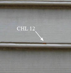 CHL 12
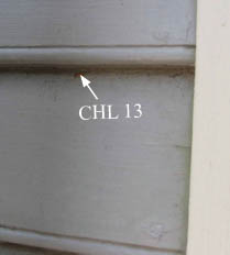 CHL 13
 CHL 19
Cornerboard samples and sample locations:
| Sample number | Sample location | Taken by/date | Samples cast | Notes |
|---|---|---|---|---|
| CHL 14* | West elevation, cornerboard on north end, in groove of bead, 16" up from bottom | N. Loeblich, July 21, 2010 | two samples cast in one resin cube, both labeled CHL 14 | |
| CHL 15 | North elevation, cornerboard on west end, paint under metal piece (possible gutter strap), nailed 6" below bed molding of cornice | N. Loeblich, July 21, 2010 | three samples cast in one resin cube, all labeled CHL 15 | |
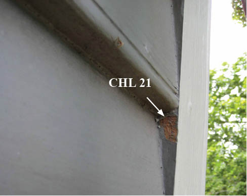
| CHL 21 | North elevation, cornerboard on west end, inner (east) edge adjoining 5th weatherboard from top | N. Loeblich, July 21, 2010 | CHL 21 | wood substrate with modern paint layers only |
Cornerboard sample locations in-situ. Photographed and sampled by N. Loeblich, July 21, 2010.
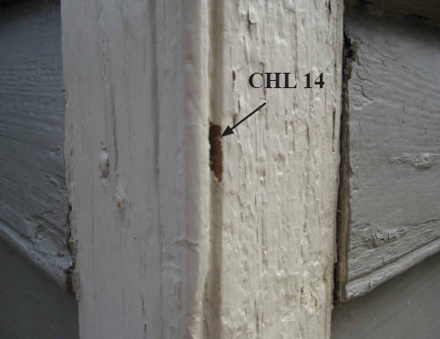 CHL 14
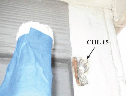 CHL 15
 CHL 21
Window frame samples and sample locations
These samples contained modern paints only and were not discussed in this report
| Sample number | Sample location | Taken by/date | Samples cast | Notes | |
|---|---|---|---|---|---|
| CHL 11 | North elevation, basement window grill, inside edge of west frame, top edge | N. Loeblich, July 21, 2010 | two samples cast in one resin cube, both labeled CHL 11 | ||
| CHL 17 | North elevation, 2nd floor, westernmost window frame, east jamb, in groove of the bead, 2' above sill | N. Loeblich, July 21, | 2010 | CHL 17 | one layer of modern paint only |
Window frame sample locations in-situ. Photographed and sampled by N. Loeblich, July 21, 2010.
 CHL 11
 CHL 17
APPENDIX B. Procedures
Sample Preparation:
The samples were cast in mini-cubes of Extec Polyester Clear Resin (methyl methacrylate monomer), polymerized with the recommended amount of methyl-ethyl-ketone catalyst. The resin was allowed to cure for 24 hours under ambient light. After cure, the individual cubes were removed from the casting tray and sanded down using a rotary sander with grits ranging from 200 — 600 to expose the cross-section surface. The samples were then dry polished with silica-embedded Micro-mesh Inc. cloths with grits ranging from 1500 to 12,000, lending the final cross-section surface a glassy-smooth finish.
Microscopy and Documentation:
The cross-section samples were examined using a Nikon Eclipse 80i microscope equipped with an EXFO X-cite 120 fluorescence illumination system fiberoptic halogen light source. Samples were examined and photographed under visible and ultraviolet light conditions (380-330 nm), at 20 to 200x magnifications. Digital images were captured using a Spot Flex digital camera with Spot Advance (version 4.6) software. All images were recorded as 12.6 MB tiff files and stored on a hard drive in a folder titled "Charlton House" on Susan Buck's laboratory computer. A separate set of images will be stored on the CWF digital database, accompanied by a digital version of the final report.
Information Provided by Visible and Ultraviolet Light Microscopy:
When examining paint cross-sections under reflected visible and ultraviolet light conditions, a number of physical characteristics can be observed to assist with the interpretation of a paint stratigraphy. These include the number and color of layers applied to a substrate, the thickness or surface texture of layers, and pigment particle size and distribution within the paint film. Relative time periods for coatings can sometimes be assigned at this stage: for instance, pre-industrial paints were hand ground, lending them a coarse, uneven surface texture with large pigment particles that vary in size and shape. By contrast, more "modern", industrially-prepared paints have smoother, even surfaces and machine-ground pigment particles of a consistent size and shape. The presence of cracks, dirt layers, or biological growth between layers can indicate presentation surfaces and/or coatings that were left exposed for an extended period of time.
Under UV light conditions, the presence and type of autofluorescence colors can distinguish sealants, clear coatings, and binding media, from darker dirt or paint layers within the stratigraphy. For instance, shellacs exhibit a distinct orange-colored autofluorescence, while natural resins (such as dammar and mastic), typically fluoresce a bright white color. Oil media tends to quench autofluorescence, while most modern, synthetic paint formulations (such as latex) exhibit no fluorescence at all. Some pigments, such as verdigris, madder, and zinc white, have distinct fluorescence characteristics, as well. UV light microscopy is critical to distinguish otherwise identical layers- such as successive varnishes, or multiple layers of whitewash.
Fluorochrome staining:
Fluorochrome stains adapted from the biological sciences were used to characterize pigments and binding media (oils, proteins, carbohydrates), in layers within the cross-section sample. The following stains were used in this analysis:
2,7 Dichlorofluorescein (DCF): 0.02% w/v in ethanol. One drop of stain was applied to the surface of the sample, blotted immediately, and cover-slipped with mineral spirits. The reaction was observed with the B-2A filter cube (450-490nm, 520 barrier filter). This stain tags for lipids, ie: oils, in a sample.
N-(-6methoxy)-8-quinoly-1-toluene sulfonamide (TSQ): 2% w/v in ethanol was dropped onto the surface of the sample, blotted, and cover-slipped with mineral spirits. The reaction was observed in ultraviolet light. This stain tags for the presence of zinc in its free ionic form, Zn+2, therefore making it an excellent marker for zinc-based pigments, which were not generally used until the introduction of zinc white (ZnO), introduced c.1835. A positive reaction is characterized by bright, sparkling bluish-white fluorescent particles in a zinc-based paint layer.
Pigment Identification with Polarized Light Microscopy:
To collect a pigment sample for polarized light microscopy (PLM), a surgical scalpel was used to collect a small scraping from a clean, representative area of paint. The blade was then pressed and pulled across a clean glass microscope slide, dispersing the pigment particles across the surface. The pigments were then permanently embedded under a cover slip using Cargille Meltmount (refractive index 1.66). The embedded pigments were then examined in cross and plane-polarized transmitted light with the Nikon Eclipse 80i microscope at 1000x magnification (using an oil immersion objective). The observed morphologies and optical properties (including color, refractive index, extinction), were compared to reference standards as well as literature sources before making final determinations.
Color measurement and matching:
Color measurements were obtained using a Minolta Chroma Meter CR-241 color analyzer/ microscope in Susan Buck's paint analysis laboratory. Equipped with an internal 360-degree pulsed xenon arc lamp, this instrument is capable of obtaining accurate color measurements in any one of five different tristimulus color measurement systems from areas as small as 0.3mm. For the purposes of this report, the CIE L*a*b* color space system (developed in 1976 by the Commission International de l'Eclairage, and now an internationally accepted industry-standard color measuring system) was used. In this system, three numerical values, known as "tristimulus" values, are obtained: L* is the lightness variable, representing dark to light on a scale of 0-100, while a* and b* are chromaticity coordinates, a* representing red to green on scale from -50 to +50, and b*representing blue to yellow on a scale from -50 to +50. These three coordinates are used to plot the location of a color in the CIE L*a*b* colorspace.
34Ideally, color measurements should be collected from a clean, unweathered sample area. If necessary, a scalpel is used to scrape an area clean before color matching. Due to inherent color variations in paints (especially in hand ground, pre-industrial coatings), three readings are taken and averaged together to establish the final CIE L*a*b* values.
These resulting values can be used to quantify color differences (ΔE), between two samples. To obtain this value, the following calculation is used:
ΔE = (ΔL*)½ + (Δa*)½ + (Δb*)½
Generally, a ΔE value ≤ 3 cannot be perceived by the human eye (Wolbers 2008). Therefore, for any two samples, ΔE values at or below this range are considered acceptable matches.
Appendix C. Sampling Memoranda
Charlton House Samples
Taken on July 20, 2010 from fragments and samples in the fragments Collection.
- CHL1 — cornice paint sample CHH-004-08, removed from north elevation cornice between 2nd and 3rd window from the west.
- CHL2 — cornice paint sample CHH-005-08, removed from north elevation cornice between 2nd and 3rd window from the west.
- CHL3 — cornice paint sample CHH-006-08, removed from north elevation cornice between 2nd and 3rd window from the west.
- CHL4 — cornice paint sample CHH-007-08, removed from north elevation cornice between 2nd and 3rd window from the west.
- CHL5 — weatherboard fragment CHH-002-08, top edge at paint ghost, 1 ¼" from right end
- CHL6 — weatherboard fragment CHH-002-08, bottom edge, underside, ½" from left end
- CHL7 — weatherboard fragment CHH-002-08, in groove of bottom edge bead, 5 ¾" from right end
Fragments Collection Items (as catalogued):
CHH-002-08 (CE-003007): Weatherboard from Charlton House: white paint (1st gen.), white paint, cream paint & varnish with poorly mixed in paint, light gray paint, white paint, gray paint (Matsen, Catherine, Cross-Section Microscopy Report, Architectural Fragments Collection, Williamsburg, VA: CWF, Architectural Research, 30 Aug. 2002. (pp. 10-12)).
Paint Samples CHH-004 through 007-08 — Removed from the North elevation of the structure from the bed molding around the modillion blocks on the cornice between the 2nd and 3rd window from the west by Tom Taylor in 1996 in preparation for repainting
- CHH-004-08 — small paint sample taken from bed molding; two samples of paint and one sample of wood
- CHH-005-08 — small rectangular paint sample taken from cornice; one section of wood with painted surface still attached
- CHH-006-08 — Small samples of paint taken from cornice; at least one sample of wood and several samples of paint
- CHH-007-08 — Small samples of paint taken from cornice; several samples of wood still have paint surface attached
To: Natasha Loeblich
From: Edward Chappell
Re: Exterior Paint Samples, Charlton House
This records where you and I took samples from the outside of Charlton House. What we observed is that the lower part of the cornice around the dentils is early pine with a substantial accumulation of paint. The corner board, weatherboards, and upper window frames include old wood, but we saw little paint accumulation. The earliest paint you saw appeared to be off-white.
(The following samples are numbered beginning with 8 due to the samples previously taken from articles in the fragments collection).
- CHL 8 — North elevation, cornice, fascia between 10th and 11th dentil from west end.
- CHL 9 — North elevation, cornice, west side of 11th dentil from west end.
- CHL10 — North elevation, cornice, fascia between 9th and 10th dentil from west end
- CHL11 — North elevation, basement window grill, inside edge of west frame, top edge
- CHL12 — North elevation, weatherboard, 6th board from the bottom, below 2nd window from west end, in groove of bead
- CHL13 — North elevation, weatherboard, 10th board up from the bottom, 4" from east pilaster of from porch, top edge
- CHL14 — West elevation, cornerboard on north end, in groove of bead, 16" up from bottom
- CHL15 — North elevation, cornerboard on west end, paint under metal piece (gutter strap?) nailed 6" below bed molding of cornice
- CHL16 — North elevation, cornice, west side of 6th dentil from west end
- CHL17 — North elevation, 2nd floor, westernmost window frame, east jamb, in groove of the bead, 2' above sill (This inner frame is more weathered than the rectangular backband, but with little paint evidence.)
- CHL18 — North elevation, face of raised fillet between 6th and 7th dentil from west end
- CHL19 — North elevation, weatherboard, 16th board from top, 1'2" east of west end, top edge immediately below upper board (Weatherboard is weathered, but looks like little paint remains.)
- CHL20 — North elevation, cornice, bottom of projecting fillet between 6th and 7th dentil from west end
- CHL21 — North elevation, cornerboard on west end, inner (east) edge adjoining 5th weatherboard from top (Not much paint)
Addendum
Analytical Report: Elemental Analysis of Charlton House Cornice Sample CHL 3b
| Subject: | Charlton House paint cross-section sample CHL 3b, taken from exterior cornice |
|---|---|
| Technique: | Scanning electron microscopy — energy dispersive spectroscopy (SEM-EDS), with backscattered imaging (SEM-BSE), and x-ray fluorescence mapping capabilities |
| Analyzed by: | Catherine Matsen, Associate Scientist, Winterthur Museum Scientific Research and Analysis Laboratory |
| Date conducted: | February 18, 2011 |
| Requested by: | Kirsten Travers, Graduate Fellow, Winterthur/University of Delaware Program in Art Conservation |
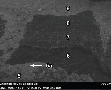
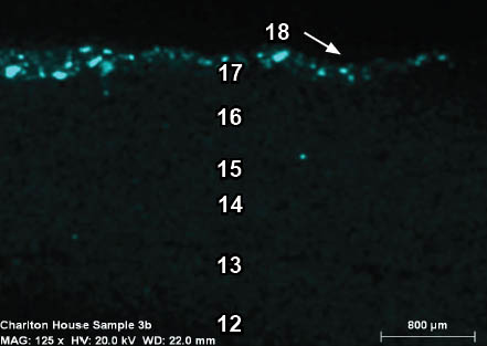
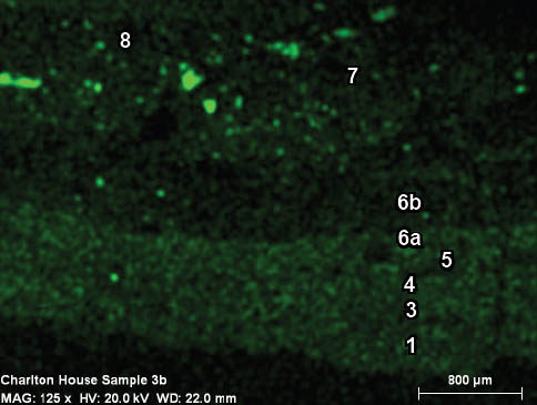
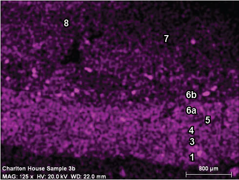
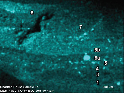
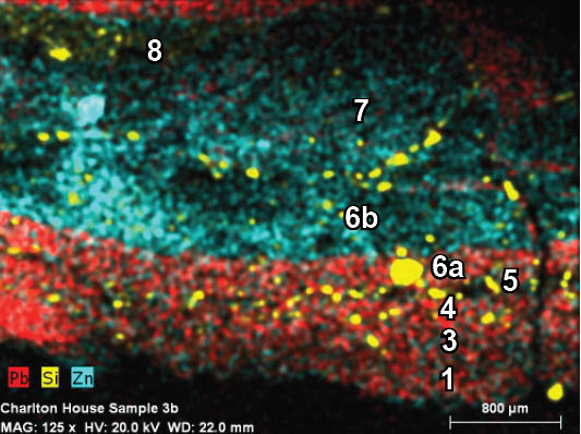
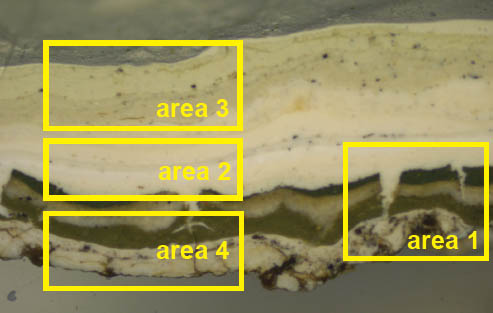 CHL 3b, visible light, 40x. The areas targeted for SEMEDS analysis are outlined in yellow
Purpose:
This analysis was carried out to determine the elemental composition of paint layers in sample CHL 3b, particularly the first paint generation. The findings will be used to determine the pigment composition of paints in the sample. This sample was one of a group analyzed by Matsen in the spring of 2011 for an ongoing study regarding the fluorescence behaviors of zinc and lead-based pigments in architectural paint cross-sections (Travers 2011).
Previous Research:
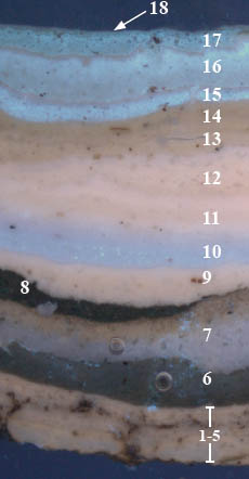
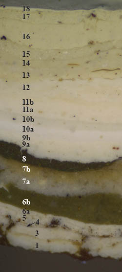
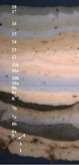
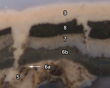
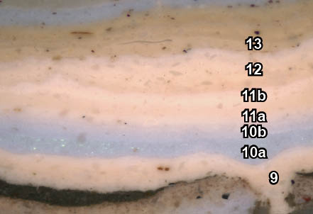
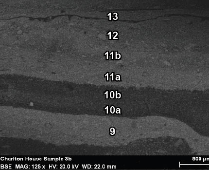
Cross-section microscopy and analysis carried out in the fall of 2010 identified eighteen finish generations in this sample, although generation 2 was determined to be missing (when compared to other Charlton House samples). The first five finish generations are white paints. The sixth paint generation consists of two layers: a white base coat and olive-green top coat. This is followed by generation 7, a gray paint with a dim autofluorescence at its surface, suggesting exposure and wear. Generation 8 is a dark green paint that exhibits a cracked and deteriorated surface. Generations 9-18 may be the primers and off-white finish coats applied by Colonial Williamsburg since 1930.
Pigment identification with polarized light microscopy (PLM), identified white lead (2PbCO3 ∙ Pb(OH)2) and chalk (CaCO3) in the first paint generation. This pigment sample was taken directly from white paints embedded deep in the wood substrate of the cornice fragment. Cross-section sample CHL 3b was stained with TSQ (N-(-6methoxy)-8-quinoly-1-toluene sulfonamide, 2% w/v in ethanol), to tag Zn+2 in the stratigraphy. Positive reactions were observed in generations 6, 7, 10, and 15-18, suggesting that zinc-based pigments such as zinc white (ZnO), or lithopone (ZnS + BaSO4), were present in these paints
Please refer to pages 8-9 of the "Cross-Section Microscopy Analysis Report: Charlton House Exterior Finishes" (Travers), for a more detailed discussion of this sample and stain reactions.
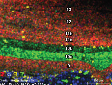
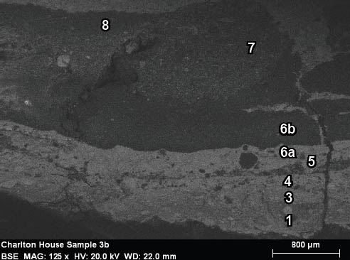
CHL 3b: labelled paint stratigraphy (in context with other Charlton House samples)
 CHL 3b, visible light, 100x
 CHL 3b, UV light, 100x
Paint generation 2 is missing from this sample.
Experimental Procedures:
The paint sample was collected from the cornice fragment with a clean scalpel blade, and stored in a small individual plastic baggie for transport to the laboratory. In the laboratory, the paint cross-section was prepared by casting the sample in a mini-cube of Extec Polyester Clear Resin (methyl methacrylate monomer), polymerized with the recommended amount of methyl ethyl ketone peroxide catalyst. The resin was allowed to cure for 24 hours under ambient light. After cure, the individual cube was removed from the casting tray and sanded down using a rotary sander with grits ranging from 200 — 600 to expose the cross-section surface. The sample was then dry polished with silica-embedded Micro-mesh Inc. cloths with grits ranging from 1500 to 12,000, lending the final cross-section surface a glassy-smooth finish, appropriate for microscopy.
The cross-section sample was examined and photographed at the paint analysis laboratory of Susan Buck, Ph.D, in Williamsburg, Virginia, using a Nikon Eclipse 80i microscope equipped with an EXFO X-cite 120 fluorescence illumination system fiberoptic halogen light source. Samples were examined and photographed under visible and ultraviolet light conditions (330-380 nm), at 40 to 400x magnifications. Fluorochrome staining was also carried out at this time. Please note that the sample was repolished to remove residual stain before it was sent to Winterthur for analysis. Digital images were captured using a Spot Flex digital camera with Spot Advance (version 4.6) software. All images were recorded as 12.6 MB tiff files and stored on a hard drive in the Charlton House file on Susan Buck's laboratory computer, and on the Colonial Williamsburg Foundation Research server with accompanying metadata.
Sample CHL 3b was sent to the Winterthur Museum Scientific and Analysis Research Laboratory (SRAL) to determine its elemental composition with scanning-electron microscopy — energy dispersive spectroscopy (SEM-EDS) with x-ray mapping and backscattered imaging capabilities. The analysis was carried out in February 2011 by Catherine Matsen, SRAL Associate Scientist.
Excess casting medium was removed with a jeweler's saw and mounted to a carbon stub with carbon tape adhesive, then coated with carbon paint up to the edge of the cross-section to prevent charge buildup. All SEM prep materials were obtained from SPI supplies. The analyses were carried out with a Topcon ABT- 60 scanning electron microscope at an accelerating voltage of 20kV at a stage height of 20mm with a 20° sample tilt in variable pressure mode to minimize charging. EDS data was analyzed with a Bruker X-flash detector and microanalysis Quantax model 200 with Esprit 1.8 software.
The data was interpreted by Kirsten Travers, WUDPAC Graduate Fellow at the Colonial Williamsburg Foundation, with the help of Catherine Matsen.
Results:
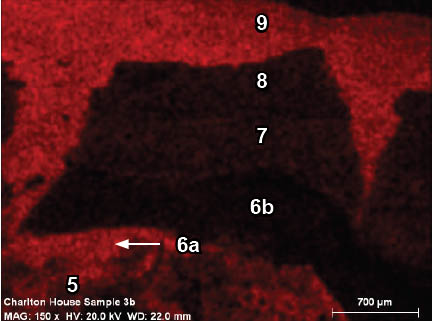
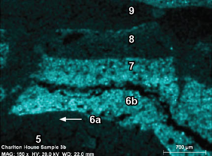
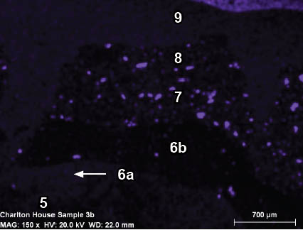
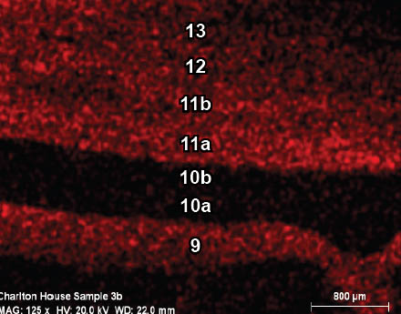
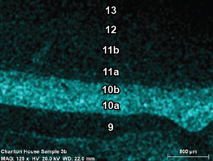
Elemental data for the entire sample was collected from four separate smaller areas (see page 1), which, when combined, gives a complete picture of the elemental composition of the sample overall. The results have been presented according to each area, starting with area 1 and ending with area 4. At the beginning of each section, the area of analysis is shown in visible light and ultraviolet light, followed by the SEMEDS backscattered image, and the individual elemental maps for every element detected in the target area, starting with the most prevalent: lead (Pb), zinc (Zn), and usually barium (Ba), or calcium (Ca). The SEM-BSE image was annotated first and these annotations were simply cut and pasted onto the elemental maps to ensure that numbering of layers was consistent. The x-ray fluorescence spectrum for each area is included at the end of each section, accompanied by a composite elemental map showing the most prominent elements present.1
All results are interpreted in a table at the end of the section, and discussed in the section that follows.
When interpreting the elemental maps, only the strongest signals, ie: solid, saturated areas of color, were considered a definite indication that an element was present. Areas of faint, inconsistent colors result from 'background noise' and do not indicate the presence of certain elements.
Overall, this analysis generated very useful data regarding the elemental composition of paints in sample CHL 3b. The majority of paints contained lead, suggesting that basic lead white is present (Pb(OH)2 ∙ 2PbCO3), although lead sulfate might also have been used (PbSO4 ∙ PbO). Other paints were rich in zinc, suggestive of zinc white (ZnO). In some instances, a combination of zinc and barium were detected, strongly indicating that lithopone (ZnS + BaSO4), was present, or a combination of zinc white (ZnO) and blanc fixe (BaSO4). Sulfur, which is present in white lead sulphate, lithopone, and blanc fixe, was not detected because this element experiences only L to K shell electron transitions (K∝ at 2.32 keV and Kb at 2.46 keV), which were overlapped by the M∝ and Mß lines of lead which occur at 2.342 keV and 2.44 keV, respectively. An instrumental technique such as Raman spectroscopy could more definitively identify these pigments in the paint cross-section.
Sample number: CHL 3b
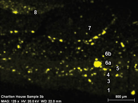
Area 1: SEM-EDS x-ray mapping results
 CHL 3b, area 1, ultraviolet light, 100x
 CHL 3b, area 1, SEM-BSE image, 150x
 CHL 3b, 150x, area 1, x-ray map for lead (Pb)
 CHL 3b, 150x, area 1, x-ray map for zinc (Zn)
 CHL 3b, 150x, area 1, x-ray map for barium (Ba)
 CHL 3b, 150x, area 1, x-ray map for aluminum (Al)
 CHL 3b, 150x, area 1, x-ray map for carbon (C)
 CHL 3b, 150x, area 1, x-ray map for calcium (Ca)
 CHL 3b, 150x, area 1, x-ray map for iron (Fe)
 CHL 3b, 150x, area 1, x-ray map for magnesium (Mg)
 CHL 3b, 150x, area 1, x-ray map for silicon (Si)
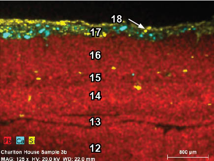
 CHL 3b, 150x, area 1, SEM-EDS x-ray map for lead (Pb), calcium (Ca), aluminum (Al), silicon (Si), zinc (Zn), iron (Fe),
and barium (Ba).
CHL 3b, 150x, area 1, SEM-EDS x-ray map for lead (Pb), calcium (Ca), aluminum (Al), silicon (Si), zinc (Zn), iron (Fe),
and barium (Ba).
An XRF spectrum was not provided for area 1.
Sample number: CHL 3b
Area 2: SEM-EDS x-ray mapping results
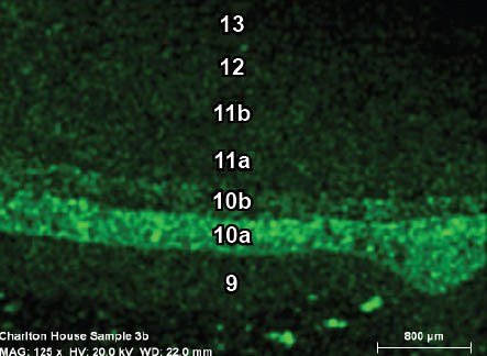
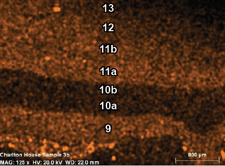
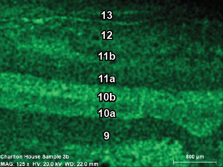
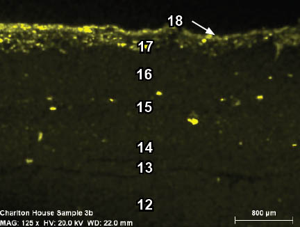
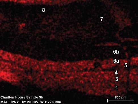
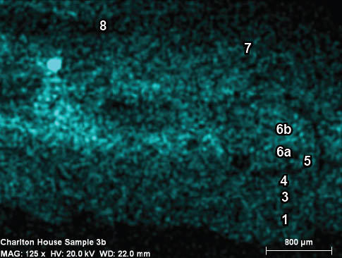
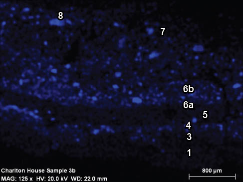
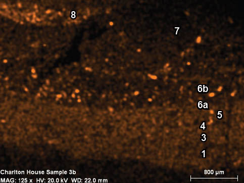
 CHL 3b, area 2, visible light, 125x
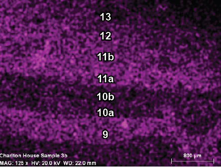
 CHL 3b, area 2, UV light, 125x
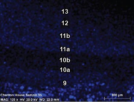
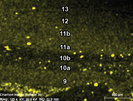
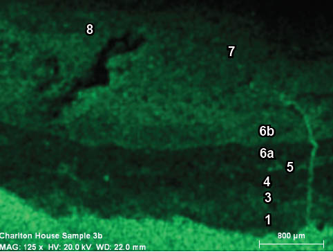
 CHL 3b, area 2, SEM-BSE image, 125x
 CHL 3b, area 2, 125x. x-ray map for lead (Pb)
 CHL 3b, area 2, 125x. x-ray map for zinc (Zn)
 CHL 3b, area 2, 125x. x-ray map for barium (Ba)
 CHL 3b, area 2, 125x. x-ray map for aluminum (Al)
 CHL 3b, area 2, 125x. x-ray map for carbon (C)
 CHL 3b, area 2, 125x. x-ray map for calcium (Ca)
 CHL 3b, area 2, 125x. x-ray map for iron (Fe)
 CHL 3b, area 2, 125x. x-ray map for potassium (K)
 CHL 3b, area 2, 125x. x-ray map for silicon (Si)
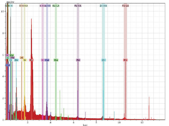
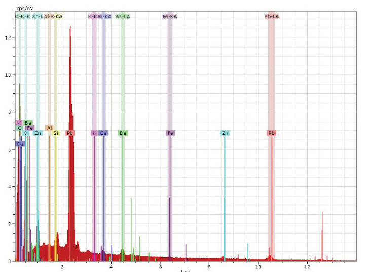 Top: SEM-EDS x-ray spectrum of sample CHL 3b, area 2. Lead (Pb), zinc (Zn), barium (Ba), calcium (Ca), and silicon (Si), are the major peaks observed.
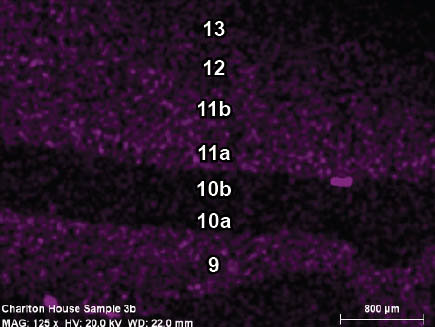
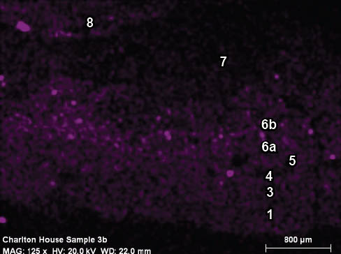
 Left: CHL 3b, 125x, area 2, SEM-EDS x-ray map for lead (Pb), calcium (Ca), silicon (Si), and barium (Ba). Zinc was not mapped
in this particular image, but was present in generations 10a and 10b.
Left: CHL 3b, 125x, area 2, SEM-EDS x-ray map for lead (Pb), calcium (Ca), silicon (Si), and barium (Ba). Zinc was not mapped
in this particular image, but was present in generations 10a and 10b.
Sample number: CHL 3b
Area 3: SEM-EDS x-ray mapping results
 CHL 3b, area 3, visible light, 125x
 CHL 3b, area 3, UV light, 125x
 CHL 3b, area 3, SEM-BSE image, 125x
 CHL 3b, area 3, SEM-EDS x-ray map for lead (Pb)
 CHL 3b, area 3, SEM-EDS x-ray map for zinc (Zn)
 CHL 3b, area 3, SEM-EDS x-ray map for titanium (Ti)
 CHL 3b, area 3, SEM-EDS x-ray map for aluminum (Al)
 CHL 3b, area 3, SEM-EDS x-ray map for calcium (Ca)
 CHL 3b, area 3, SEM-EDS x-ray map for magnesium (Mg)
 CHL 3b, area 3, SEM-EDS x-ray map for silicon (Si)
 CHL 3b, area 3, SEM-EDS x-ray map for lead (Pb), calcium (Ca), titanium (Ti), carbon (C)
 CHL 3b, area 3, SEM-EDS x-ray map for lead (Pb), calcium (Ca), silicon (Si)
 Top: SEM-EDS x-ray spectrum of sample CHL 3b, area 3. Lead (Pb), zinc (Zn), titanium (Ti), calcium (Ca), and silicon (Si), are
the major peaks observed.
Top: SEM-EDS x-ray spectrum of sample CHL 3b, area 3. Lead (Pb), zinc (Zn), titanium (Ti), calcium (Ca), and silicon (Si), are
the major peaks observed.
 Left: CHL 3b, 125x, area 3, SEM-EDS x-ray map for lead (Pb), zinc (Zn), and calcium (Ca).
Sample number: CHL 3b
Area 4: SEM-EDS x-ray mapping results
 CHL 3b, area 4, visible light, 125x
 CHL 3b, area 4, SEM-BSE image, 125x
 CHL 3b, area 4, SEM-EDS x-ray map for lead (Pb)
 CHL 3b, area 4, SEM-EDS x-ray map for zinc (Zn)
 CHL 3b, area 4, SEM-EDS x-ray map for calcium (Ca)
 CHL 3b, area 4, SEM-EDS x-ray map for barium (Ba)
 CHL 3b, area 4, SEM-EDS x-ray map for aluminum (Al)
 CHL 3b, area 4, SEM-EDS x-ray map for carbon (C)
 CHL 3b, area 4, SEM-EDS x-ray map for iron (Fe)
 CHL 3b, area 4, SEM-EDS x-ray map for potassium (K)
 CHL 3b, area 4, SEM-EDS x-ray map for oxygen (O)
 CHL 3b, area 4, SEM-EDS x-ray map for silicon (Si)
 Top: SEM-EDS x-ray spectrum of sample CHL 3b, area 4. Lead (Pb), zinc (Zn), barium (Ba), calcium (Ca), and silicon (Si),
are the major peaks observed.
Top: SEM-EDS x-ray spectrum of sample CHL 3b, area 4. Lead (Pb), zinc (Zn), barium (Ba), calcium (Ca), and silicon (Si),
are the major peaks observed.
 Left: CHL 3b, 125x, area 4, SEM-EDS x-ray map for lead (Pb), zinc (Zn), and silicon (Si).
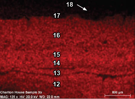
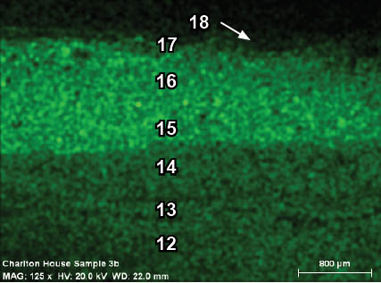
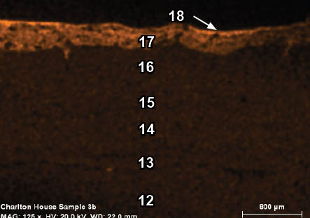
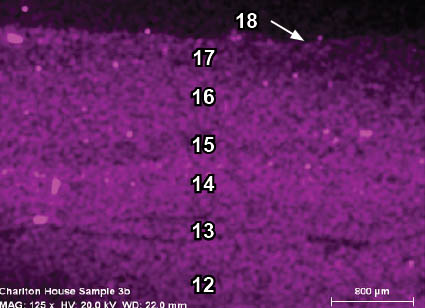
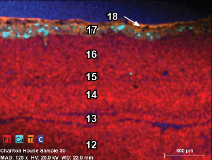
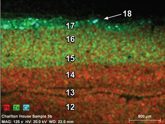
| Generation | appearance in visible light | appearance in ultraviolet light | elements detected | interpretation |
|---|---|---|---|---|
| 18 | cream color | gray, dim | Ti, Ca, Mg, Si | Titanium white (TiO2), with chalk (CaCO3), silicate (SiO2), or talc (3MgO ∙ 4SiO2 ∙ H2O), fillers |
| 17 | cream color | dim bluish | Zn, Ti | zinc white (ZnO), titanium white (TiO2) |
| 16 | cream color | dim, blue with twinkling particles present | Pn, Zn | lead white (Pb(OH)2∙2Pb(CO)3), and zinc white (ZnO) |
| 15 | cream color | light blue | Pb, Zn | lead white (Pb(OH)2∙2Pb(CO)3), and zinc white (ZnO) |
| 14 | cream color | pinkish | Pb | lead white (Pb(OH)2∙2Pb(CO)3) |
| 13 | cream color | pinkish | Pb | lead white (Pb(OH)2∙2Pb(CO)3) |
| 12 | cream color | pinkish | Pb | lead white (Pb(OH)2∙2Pb(CO)3) |
| 11 | cream color | pinkish | Pb | lead white (Pb(OH)2∙2Pb(CO)3) |
| 10b | light gray | light blue, faint twinkling particles | Zn, Ba, Si | lithopone (ZnS + BaSO4), or mixture of zinc white (ZnO) and blanc fixe (BaSO4), Si from quartz (SiO2) or polishing cloths |
| 10a | white | light blue, bright twinkling particles | Zn, Ba, Si | lithopone (ZnS + BaSO4), or mixture of zinc white (ZnO) and blanc fixe (BaSO4), Si from quartz (SiO2) or polishing cloths |
| 9 | cream color | pinkish | Pb | lead white (Pb(OH)2∙2Pb(CO)3) |
| 8 | dark green | none | Fe, Al, Si, Ca | iron earths (Fe2O3 ∙ nH2O), aluminosilicate clay inclusions, chalk (CaCO3) |
| 7 | gray | dim, gray faint twinkling particles | Zn, O, K, Ba, Ca, Fe, Al | zinc white (ZnO), K from organic media, chalk (CaCO3), Ba, Fe, Al earth minerals in chalk (not enough Ba for lithopone or blanc fixe) |
| 6b | olive-green | dim, faint twinkling particles | Zn, Ca, Al, K, Si | zinc white (ZnO), chalk (CaCO3), K from organic media, Al and Si from aluminosilicate clays |
| 6a | cream color | pinkish | Pb, K, O, Si | lead white (Pb(OH)2∙2Pb(CO)3), K from organic media, quartz (SiO2) or Si from polishing cloth |
| 5 | cream color | pinkish | Pb, K, O, Si | lead white (Pb(OH)2∙2Pb(CO)3), K from organic media, quartz (SiO2) or Si from polishing cloth |
| 4 | cream color | pinkish | Pb, K, Ca, O, Si | lead white (Pb(OH)2∙2Pb(CO)3), K from organic media, chalk (CaCO3), quartz (SiO2) or Si from polishing cloth |
| 3 | cream color | pinkish | Pb, K | lead white (Pb(OH)2∙2Pb(CO)3), K from organic media |
| 2 | missing | n/a | n/a | n/a |
| 1 | cream color | pinkish | Pb, K | lead white (Pb(OH)2∙2Pb(CO)3), K from organic media |
Discussion:
The two most dominant elements in sample CHL 3b were lead (Pb), and zinc (Zn). This can be interpreted as basic lead white (2PbCO3 role="label" Pb(OH)2), and zinc white (ZnO), and/or lithopone (ZnS + BaSO4), or a combination of zinc white and blanc fixe (BaSO4), all of which are white pigments commonly found in housepaints. Basic lead white has been used from antiquity to the present day, so the presence of this pigment could not be used to assign a particular date to paint layers. However, zinc-based pigments such as zinc white and lithopone were not commercially introduced to the housepainting market until the nineteenth century (c.1845 and c.1870, respectively). Paints which contain zinc would therefore post-date the colonial period, making these results extremely relevant in the assignment of paint layers to the 18th century. In consideration of this evidence, it can be said with certainty that generations 6b and later postdate the colonial period. Generation 10 appears to contain a mixture of zinc and barium, strongly suggesting that lithopone is present. Therefore, these layers would post-date c.1870. The presence of titanium (Ti) in generations 17 and 18 strongly suggest that titanium white (TiO2), is present. This pigment was introduced c.1915 and has largely superceded lead and zinc-based white pigments in modern paint media, suggesting a 20th-century date for these paints. Furthermore, the dim autofluorescence of these generations indicates a synthetic binding media, suggesting that these paints could have been applied anywhere from the mid-20th-century to the present day.
The presence of potassium (K) in many of the paint layers can be attributed to organic media, such as linseed oil (Matsen 2011, pers. comm.). Calcium (Ca) could be attributed to calcium carbonate, or chalk (CaCO3), a common extender added to housepaints. Likewise, aluminum (Al), and silica (Si), suggest the presence of aluminosilicate clays that may have been added as fillers, or inadvertent inclusions accompanying earth pigments such as ochres, or chalk. Furthermore, it is important to note that the cross-sections were polished with silica-embedded cloths, which could also account for the presence of silica in the spectra.
Conclusion:
The analytical results provided critical evidence regarding relative dates for paint layers and the elemental composition of the first generation paint in Charlton House sample CHL3b, from which the pigments could be interpreted.
The first paint generation contains mostly lead (Pb), strongly suggesting that basic lead white (2PbCO3 role="label" Pb(OH)2) is present. This pigment has been in use since antiquity, and was a ubiquitous housepainting pigment up to the mid-late 20th-century in the United States, so its presence does not provide a date for generation 1. However, this analysis lends support to the PLM findings which determined the first generation paint contained lead white. While PLM also detected chalk (CaCO3), no calcium (Ca) was detected in generation 1 with SEM-EDS x-ray mapping. One possible explanation for this discrepancy is that since the pigment sample for PLM was collected from a cream paint embedded deep in the wood cells of the cornice substrate, it is possible that the pigments did not belong to the first generation, but might have been generation 4, a cream-colored paint that was found to contain both lead and calcium with this analysis.
Additional techniques such as Raman spectroscopy or x-ray diffraction spectroscopy (XRD), should be carried out to conclusively identify basic lead white, and to search for any evidence of chalk in the first generation.
 CHL 1a VIS 40x
 CHL 1a UV 40x
 CHL 2a VIS 20x
 CHL 2a UV 20x
 CHL 2a VIS 40x
 CHL 2a UV 40x
 CHL 2a VIS 100x
 CHL 2a UV 100x
 CHL 2a UV b4TSQ 100x
 CHL 2a UV TSQ 100x
 CHL 2b VIS 100x
 CHL 2b UV 100x
 CHL 2b UV b4TSQ 100x
 CHL 2b UV TSQ 100x
 CHL 3a VIS 100x
 CHL 3a UV 100x
 CHL 3a VIS 200x
 CHL 3a UV 200x
 CHL 3a UV b4TSQ 200x
 CHL 3a UV TSQ 200x
 CHL 3a B2A b4DCF 200x
 CHL 3a B2A DCF 200x
 CHL 3b 2 layer 1 BF 1000x
 CHL 3b 2 layer 1 DF 1000x
 CHL 3b layer 1 BF 100x
 CHL 3b layer 1 DF 100x
 CHL 3b VIS 40x
 CHL 3b UV 40x
 CHL 3b UV b4TSQ 40x
 CHL 3b UV TSQ 40x
 CHL 3b VIS2 100x
 CHL 3b UV1 100x
 CHL 3b VIS1 100x
 CHL 3b UV2 100x
 CHL 4a VIS 200x
 CHL 4a UV 200x
 CHL 4a UV b4TSQ 200x
 CHL 4a UV TSQ 200x
 CHL 4b VIS 40x
 CHL 4b UV 40x
 CHL 5a VIS 100x
 CHL 5a UV 100x
 CHL 5a VIS 200x
 CHL 5a UV 200x
 CHL 5a B2A b4DCF 100x
 CHL 5a B2A DCF 100x
 CHL 5b VIS2 100x
 CHL 5b UV1 100x
 CHL 5b VIS1 100x
 CHL 5b UV2 100x
 CHL 6 VIS 100x
 CHL 6 UV 100x
 CHL 6 VIS2 100x
 CHL 6 UV2 100x
 CHL 7 VIS 100x
 CHL 7 UV 100x
 CHL 8 VIS 40x
 CHL 8 UV 40x
 CHL 8 VIS2 100x
 CHL 8 UV2 100x
 CHL 8 VIS 3 100x
 CHL 8 UV3 100x
 CHL 9 resanded VIS 100x
 CHL 9 resanded UV 100x
 CHL 9 wood VIS 100x
 CHL 9 wood UV 100x
 CHL 10 paint VIS 100x
 CHL 10 paint UV 100x
 CHL 10 B2A b4DCF 100x
 CHL 10 B2A DCF 100x
 CHL 10 wood VIS 100x
 CHL 10 wood UV 100x
 CHL 11 VIS 100x
 CHL 11 UV 100x
 CHL 11 VIS 200x
 CHL 11 UV 200x
 CHL 11 UV b4TSQ 200x
 CHL 11 UV TSQ 200x
 CHL 11 wood VIS 200x
 CHL 11 wood UV 200x
 CHL 12 paint VIS 100x
 CHL 12 paint UV 100x
 CHL 12 VIS 100x
 CHL 12 UV 100x
 CHL 12 wood VIS 100x
 CHL 12 wood UV 100x
 CHL 13 paint VIS 100x
 CHL 13 paint UV 100x
 CHL 13 wood VIS 200x
 CHL 13 wood UV 100x
 CHL 13 wood UV b4TSQ 200x
 CHL 13 wood UV TSQ 200x
 CHL 14 paint 2 VIS 200x
 CHL 14 paint 2 UV 200x
 CHL 14 paint VIS 100x
 CHL 14 paint UV 100x
 CHL 14 wood VIS 100x
 CHL 14 wood UV 100x
 CHL 14 wood2 VIS 200x
 CHL 14 wood2 UV 200x
 CHL 14 wood2 UV b4DCF 200x
 CHL 14 wood2 UV DCF 200x
 CHL 15 VIS 100x
 CHL 15 UV 100x
 CHL 15 VIS 200x
 CHL 15 UV 200x
 CHL 15b VIS1 100x
 CHL 15b UV1 100x
 CHL 15b VIS2 100x
 CHL 15b UV2 100x
 CHL 15b VIS3 100x
 CHL 15b UV3 100x
 CHL 16 VIS 100x
 CHL 16 UV 100x
 CHL 16 VIS2 100x
 CHL 16 UV2 100x
 CHL 17 VIS 100x
 CHL 17 UV 100x
 CHL 18 VIS 40x
 CHL 18 UV 40x
 CHL 18 VIS 100x
 CHL 18 UV 100x
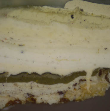 CHL 18 VIS2 100x
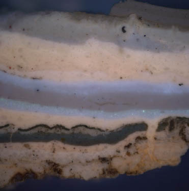 CHL 18 UV2 100x
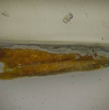 CHL 19 VIS 40x
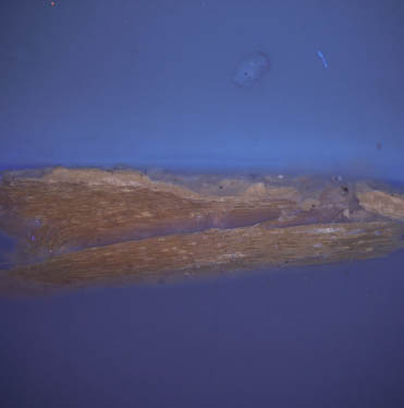 CHL 19 UV 40x
 CHL 19 VIS 200x
 CHL 19 UV 200x
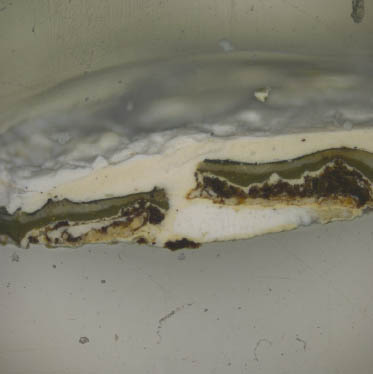 CHL 20 VIS 40x
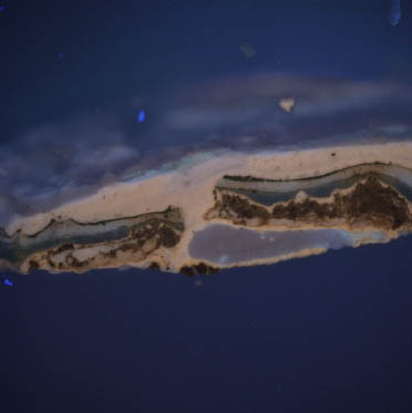 CHL 20 UV 40x
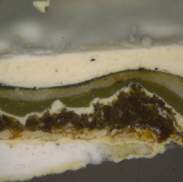 CHL 20 VIS 100x
 CHL 20 UV 100x
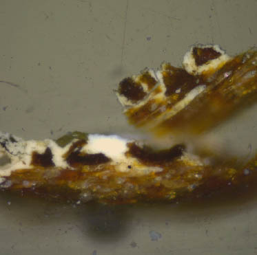 CHL 20 wood VIS 100x
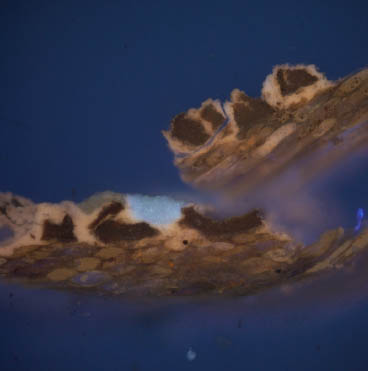 CHL 20 wood UV 100x
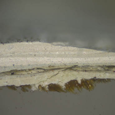 CHL 21 VIS 100x
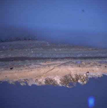 CHL 21 UV 100x
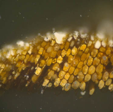 CHL1b resanded VIS 200x
 CHL1b resanded UV 200x
 CHL1b resanded UV b4 TSQ 200x
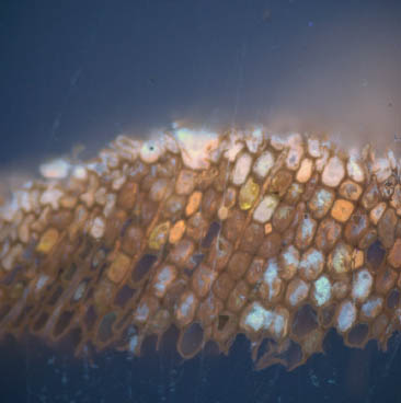 CHL1b resanded UV TSQ 200x
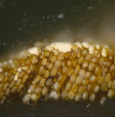 CHL1b VIS 200x
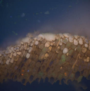 CHL1b UV 200x
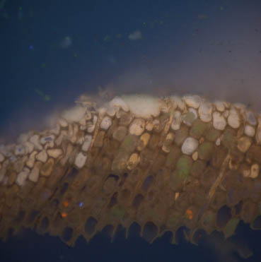 CHL1b UV b4 DCF 200x
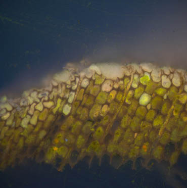 CHL1b UV DCF 200x
 weatherboard white BF 400x
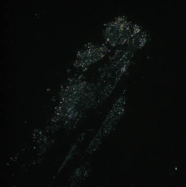 weatherboard white DF 400x
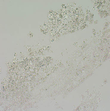 weatherboard white BF 1000x
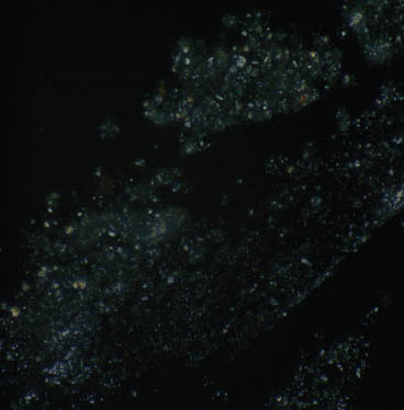 weatherboard white DF 1000x